Tuesday Poster Session
Category: Interventional Endoscopy
P3749 - Diagnosis of Recurrent Ewing Sarcoma With Endoscopic Ultrasound-Guided Fine Needle Aspiration of a Lung Mass: A Case Report
Tuesday, October 24, 2023
10:30 AM - 4:00 PM PT
Location: Exhibit Hall


Natalie Wilson, MD
University of Minnesota
Minneapolis, MN
Presenting Author(s)
Natalie Wilson, MD1, Abdulfatah Issak, MD2, Khalid Amin, MD2, Guru Trikudanathan, MD2, Shawn Mallery, MD2
1University of Minnesota, Minneapolis, MN; 2University of Minnesota Medical Center, Minneapolis, MN
Introduction: Endoscopic ultrasound-guided fine needle aspiration (EUS-FNA) is commonly used to sample lesions in the gastrointestinal tract. It has infrequently been described as a modality to sample and diagnose pulmonary lesions. Here, we describe a case of a patient with recurrent Ewing sarcoma that was diagnosed with EUS-FNA after presenting with a lung mass.
Case Description/Methods: A 28-year-old female with a history of cystic fibrosis, breast cancer status post bilateral mastectomy and chemotherapy, and Ewing sarcoma of the left tibia diagnosed 13 years ago and treated with surgical excision and chemotherapy, was discovered to have a lung mass on CT imaging obtained for breast cancer surveillance. Imaging revealed a 4.7 cm paraesophageal mediastinal mass. Given the location of the mass, she was referred for EUS-FNA.
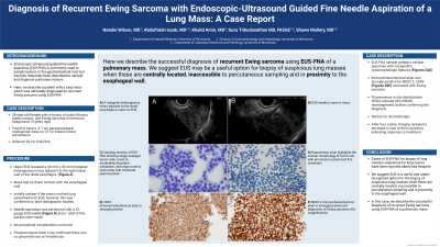
Upper EUS revealed a triangular heterogenous mass adjacent to the right lateral wall of the distal esophagus measuring 40 mm x 36 mm [Figure 1A]. There was no direct contact with the esophageal wall. It was unclear if the mass involved the lung parenchyma based on EUS, however, this was confirmed on later radiographic studies. Fine needle aspiration was performed with a 25-gauge EchoTip endoscopic ultrasound needle [Figure 1B]. A postprocedural chest x-ray confirmed there was no pneumothorax or hemothorax.
The EUS-FNA sample yielded a cellular specimen with nonspecific cytomorphologic features [Figure 1 C&D]. Immunohistochemical stains showed tumor cells that were strongly positive for NKX2.2 and CD99 [Figure 1 E&F] consistent with Ewing sarcoma. Fluorescence in situ hybridization (FISH) showed 93% EWSR1 rearrangement, further confirming the diagnosis. The patient was started on chemotherapy. After four cycles, imaging revealed a decrease in size of the lung lesion, indicating response to treatment.
Discussion: In this case, we describe the successful diagnosis of recurrent Ewing sarcoma using EUS-FNA of a pulmonary mass. No procedure-related adverse events occurred. Cases of EUS-FNA for biopsy of lung masses suspicious for lung cancer have been reported albeit less frequent. We suggest EUS is a safe, useful, and under-recognized option for the biopsy of suspicious lung masses when these are centrally located, inaccessible to percutaneous sampling, and in proximity to the esophageal wall such as in this case.

Disclosures:
Natalie Wilson, MD1, Abdulfatah Issak, MD2, Khalid Amin, MD2, Guru Trikudanathan, MD2, Shawn Mallery, MD2. P3749 - Diagnosis of Recurrent Ewing Sarcoma With Endoscopic Ultrasound-Guided Fine Needle Aspiration of a Lung Mass: A Case Report, ACG 2023 Annual Scientific Meeting Abstracts. Vancouver, BC, Canada: American College of Gastroenterology.
1University of Minnesota, Minneapolis, MN; 2University of Minnesota Medical Center, Minneapolis, MN
Introduction: Endoscopic ultrasound-guided fine needle aspiration (EUS-FNA) is commonly used to sample lesions in the gastrointestinal tract. It has infrequently been described as a modality to sample and diagnose pulmonary lesions. Here, we describe a case of a patient with recurrent Ewing sarcoma that was diagnosed with EUS-FNA after presenting with a lung mass.
Case Description/Methods: A 28-year-old female with a history of cystic fibrosis, breast cancer status post bilateral mastectomy and chemotherapy, and Ewing sarcoma of the left tibia diagnosed 13 years ago and treated with surgical excision and chemotherapy, was discovered to have a lung mass on CT imaging obtained for breast cancer surveillance. Imaging revealed a 4.7 cm paraesophageal mediastinal mass. Given the location of the mass, she was referred for EUS-FNA.
Upper EUS revealed a triangular heterogenous mass adjacent to the right lateral wall of the distal esophagus measuring 40 mm x 36 mm [Figure 1A]. There was no direct contact with the esophageal wall. It was unclear if the mass involved the lung parenchyma based on EUS, however, this was confirmed on later radiographic studies. Fine needle aspiration was performed with a 25-gauge EchoTip endoscopic ultrasound needle [Figure 1B]. A postprocedural chest x-ray confirmed there was no pneumothorax or hemothorax.
The EUS-FNA sample yielded a cellular specimen with nonspecific cytomorphologic features [Figure 1 C&D]. Immunohistochemical stains showed tumor cells that were strongly positive for NKX2.2 and CD99 [Figure 1 E&F] consistent with Ewing sarcoma. Fluorescence in situ hybridization (FISH) showed 93% EWSR1 rearrangement, further confirming the diagnosis. The patient was started on chemotherapy. After four cycles, imaging revealed a decrease in size of the lung lesion, indicating response to treatment.
Discussion: In this case, we describe the successful diagnosis of recurrent Ewing sarcoma using EUS-FNA of a pulmonary mass. No procedure-related adverse events occurred. Cases of EUS-FNA for biopsy of lung masses suspicious for lung cancer have been reported albeit less frequent. We suggest EUS is a safe, useful, and under-recognized option for the biopsy of suspicious lung masses when these are centrally located, inaccessible to percutaneous sampling, and in proximity to the esophageal wall such as in this case.

Figure: Figure 1.
A) A triangular heterogenous mass adjacent to the distal esophagus is seen on EUS
B) Needle is seen in mass
C) Cytology smears of EUS-FNA showing singly arranged tumor cells, scant to moderately abundant cytoplasm, and large round to oval nuclei with moderate pleomorphism
D) Papaniclaou stain highlights the nuclear morphology of tumor cell with prominent nucleoli and fine chromatin
E) CD99 immunohistochemical stain is strongly positive
F) NKX2.2 immunohistochemical stain is strongly positive and diagnostic of Ewing sarcoma (40x magnification)
Disclosures:
Natalie Wilson indicated no relevant financial relationships.
Abdulfatah Issak indicated no relevant financial relationships.
Khalid Amin indicated no relevant financial relationships.
Guru Trikudanathan: Boston Scientific Coporation – Consultant.
Shawn Mallery indicated no relevant financial relationships.
Natalie Wilson, MD1, Abdulfatah Issak, MD2, Khalid Amin, MD2, Guru Trikudanathan, MD2, Shawn Mallery, MD2. P3749 - Diagnosis of Recurrent Ewing Sarcoma With Endoscopic Ultrasound-Guided Fine Needle Aspiration of a Lung Mass: A Case Report, ACG 2023 Annual Scientific Meeting Abstracts. Vancouver, BC, Canada: American College of Gastroenterology.
