Tuesday Poster Session
Category: Colon
P3070 - Primary Gastrointestinal Tract Melanoma with Metastatic Spread: A Case Series Illuminating Diagnostic Challenges and Prognostic Considerations
Tuesday, October 24, 2023
10:30 AM - 4:00 PM PT
Location: Exhibit Hall

Has Audio
- KT
Kelsey Theriault, DO
Florida State University
Cape Coral, FL
Presenting Author(s)
Gautam Maddineni, MD, Kelsey Theriault, DO, Augustine Salami, MD, Katarzyna Karpinska Leydier, MD, Stefan Multinovic, MD, Edward Jurkovic, DO
Florida State University, Cape Coral, FL
Introduction: Primary gastrointestinal (GI) tract melanoma is a rare condition. This case series presents two cases of primary GI tract melanoma with metastatic spread, highlighting the need for a better understanding of risk factors and long-term survival rates associated with this disease.
Case Description/Methods: Case 1:
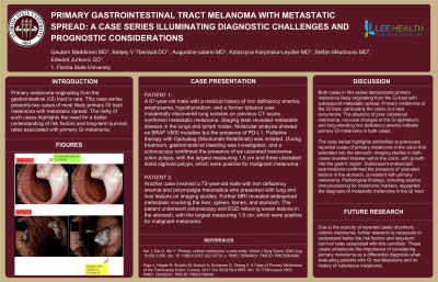
A 67-year-old male with a medical history of iron deficiency anemia, emphysema, hypothyroidism, and a former tobacco user. Incidentally discovered lung nodules on previous CT scans confirmed metastatic melanoma. Staging tests revealed metastatic disease in the lungs and lymph nodes. Molecular analysis showed no BRAF V600 mutation but the presence of PD-L1. Palliative therapy with Opdualag (Nivolumab-Relatlimab) was initiated. During treatment, gastrointestinal bleeding was investigated, and a colonoscopy confirmed the presence of six ulcerated transverse colon polyps, with the largest measuring 1.5 cm (IMAGE) and three ulcerated distal sigmoid polyps, which were positive for malignant melanoma.
Case 2:
Another case involved a 73-year-old male with iron deficiency anemia and polymyalgia rheumatica who presented with lung and liver lesions on imaging studies. Further MRI revealed widespread metastasis involving the liver, spleen, bones, and stomach. The patient underwent colonoscopy and EGD relieving seven lesions in the stomach, with the largest measuring 1.5 cm (IMAGE), which were positive for malignant melanoma.
Discussion: Both cases demonstrate likely primary melanoma originating from the GI tract with subsequent metastatic spread. Primary GI melanoma, particularly in the colon, is rare. The absence of prior cutaneous melanoma, mucosal changes in the GI tract, and long-standing iron deficiency anemia suggest primary GI melanoma in both cases.
These cases resemble previously reported primary melanoma in the colon extending into the stomach. Imaging studies and endoscopic examinations confirmed the presence of masses and ulcerated lesions in the colon and stomach, respectively. Positive immunostaining for melanoma markers supported the diagnosis of melanoma in the GI tract.
Given the limited number of reported cases of primary colonic melanoma, further research is needed to understand the risk factors and long-term survival rates associated with this condition. These cases emphasize the importance of considering primary melanoma as a possible diagnosis when evaluating patients with GI manifestations and no history of cutaneous melanoma.

Disclosures:
Gautam Maddineni, MD, Kelsey Theriault, DO, Augustine Salami, MD, Katarzyna Karpinska Leydier, MD, Stefan Multinovic, MD, Edward Jurkovic, DO. P3070 - Primary Gastrointestinal Tract Melanoma with Metastatic Spread: A Case Series Illuminating Diagnostic Challenges and Prognostic Considerations, ACG 2023 Annual Scientific Meeting Abstracts. Vancouver, BC, Canada: American College of Gastroenterology.
Florida State University, Cape Coral, FL
Introduction: Primary gastrointestinal (GI) tract melanoma is a rare condition. This case series presents two cases of primary GI tract melanoma with metastatic spread, highlighting the need for a better understanding of risk factors and long-term survival rates associated with this disease.
Case Description/Methods: Case 1:
A 67-year-old male with a medical history of iron deficiency anemia, emphysema, hypothyroidism, and a former tobacco user. Incidentally discovered lung nodules on previous CT scans confirmed metastatic melanoma. Staging tests revealed metastatic disease in the lungs and lymph nodes. Molecular analysis showed no BRAF V600 mutation but the presence of PD-L1. Palliative therapy with Opdualag (Nivolumab-Relatlimab) was initiated. During treatment, gastrointestinal bleeding was investigated, and a colonoscopy confirmed the presence of six ulcerated transverse colon polyps, with the largest measuring 1.5 cm (IMAGE) and three ulcerated distal sigmoid polyps, which were positive for malignant melanoma.
Case 2:
Another case involved a 73-year-old male with iron deficiency anemia and polymyalgia rheumatica who presented with lung and liver lesions on imaging studies. Further MRI revealed widespread metastasis involving the liver, spleen, bones, and stomach. The patient underwent colonoscopy and EGD relieving seven lesions in the stomach, with the largest measuring 1.5 cm (IMAGE), which were positive for malignant melanoma.
Discussion: Both cases demonstrate likely primary melanoma originating from the GI tract with subsequent metastatic spread. Primary GI melanoma, particularly in the colon, is rare. The absence of prior cutaneous melanoma, mucosal changes in the GI tract, and long-standing iron deficiency anemia suggest primary GI melanoma in both cases.
These cases resemble previously reported primary melanoma in the colon extending into the stomach. Imaging studies and endoscopic examinations confirmed the presence of masses and ulcerated lesions in the colon and stomach, respectively. Positive immunostaining for melanoma markers supported the diagnosis of melanoma in the GI tract.
Given the limited number of reported cases of primary colonic melanoma, further research is needed to understand the risk factors and long-term survival rates associated with this condition. These cases emphasize the importance of considering primary melanoma as a possible diagnosis when evaluating patients with GI manifestations and no history of cutaneous melanoma.

Figure: Colonoscopy and Endoscopy finding images
Disclosures:
Gautam Maddineni indicated no relevant financial relationships.
Kelsey Theriault indicated no relevant financial relationships.
Augustine Salami indicated no relevant financial relationships.
Katarzyna Karpinska Leydier indicated no relevant financial relationships.
Stefan Multinovic indicated no relevant financial relationships.
Edward Jurkovic indicated no relevant financial relationships.
Gautam Maddineni, MD, Kelsey Theriault, DO, Augustine Salami, MD, Katarzyna Karpinska Leydier, MD, Stefan Multinovic, MD, Edward Jurkovic, DO. P3070 - Primary Gastrointestinal Tract Melanoma with Metastatic Spread: A Case Series Illuminating Diagnostic Challenges and Prognostic Considerations, ACG 2023 Annual Scientific Meeting Abstracts. Vancouver, BC, Canada: American College of Gastroenterology.
