Tuesday Poster Session
Category: Biliary/Pancreas
P2904 - What Made the Gallbladder Angry? A Case of Ipilimumab and Nivolumab Induced Acute Cholecystitis
Tuesday, October 24, 2023
10:30 AM - 4:00 PM PT
Location: Exhibit Hall

Has Audio

Balakrishna Ravella, MBBS, MD
OSF Health St. Mary's Medical Center
Galesburg, IL
Presenting Author(s)
Balakrishna Ravella, MBBS, MD1, Sana Pirzada, MD2, Mikala Brinkman, MD1, Afshin Khaiser, MD1
1OSF Health St. Mary's Medical Center, Galesburg, IL; 2St. Luke's Hospital, Chesterfield, MO
Introduction: The recent advent of Immune checkpoint inhibitors (ICIs) has revolutionized the cancer treatment. Despite improvement in clinical oncologic endpoints, ICIs are known to cause several immune-related adverse events (irAEs), especially when used in combination. We present a patient who developed acute cholecystitis after initiating ipilimumab and nivolumab for metastatic malignant melanoma.
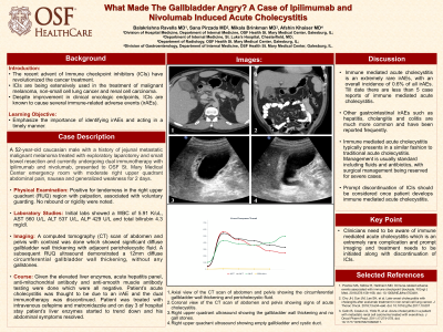
Case Description/Methods: A 52-year-old male with a history of jejunal metastatic malignant melanoma treated with exploratory laparotomy and small bowel resection and currently undergoing immunotherapy with ipilimumab and nivolumab, presented to our emergency department with right upper quadrant abdominal pain and nausea for 2 days. His last known cycle of immunotherapy was 2 weeks prior to this presentation. Labs showed a WBC of 5.91K/uL, AST 560 U/L, ALT 537 U/L, ALP 429 U/L and total bilirubin 4.3 mg/dl. A computed tomography (CT) scan of abdomen and pelvis with contrast was done which showed diffuse gallbladder wall thickening with adjacent pericholecystic fluid (Figure 1&2). Subsequently a right upper quadrant ultrasound was also done which again showed a 12 mm diffuse circumferential gallbladder wall thickening, without any associated gallstones (Figure 3&4). A decision was made to treat the patient conservatively and was started on 0.9% normal saline, intravenous metronidazole and cefepime. Patient’s liver enzymes eventually trended down (Table 1) and his right upper quadrant abdominal pain improved. At the follow up visit after discharge, patient’s abdominal pain was completely resolved, however the combination immunotherapy was discontinued given grade 3 transaminitis and acute cholecystitis.
Discussion: Immune mediated acute cholecystitis (IAC) is an extremely rare irAEs, with an overall incidence of 0.6% of all irAEs. Till date there are less than 5 case reports of immune mediated acute cholecystitis. Other gastrointestinal irAEs such as hepatitis, cholangitis and colitis are much more common and have been reported frequently. IAC typically presents in a similar fashion to traditional acute cholecystitis. Management is usually standard including fluids and antibiotics, and surgical management being reserved for severe cases. However, prompt discontinuation of ICIs should be considered once patient develops IAC.
In conclusion, clinicians need to be aware of IAC which is an extremely rare complication and prompt imaging, and treatment needs to be initiated along with discontinuation of ICIs.

Disclosures:
Balakrishna Ravella, MBBS, MD1, Sana Pirzada, MD2, Mikala Brinkman, MD1, Afshin Khaiser, MD1. P2904 - What Made the Gallbladder Angry? A Case of Ipilimumab and Nivolumab Induced Acute Cholecystitis, ACG 2023 Annual Scientific Meeting Abstracts. Vancouver, BC, Canada: American College of Gastroenterology.
1OSF Health St. Mary's Medical Center, Galesburg, IL; 2St. Luke's Hospital, Chesterfield, MO
Introduction: The recent advent of Immune checkpoint inhibitors (ICIs) has revolutionized the cancer treatment. Despite improvement in clinical oncologic endpoints, ICIs are known to cause several immune-related adverse events (irAEs), especially when used in combination. We present a patient who developed acute cholecystitis after initiating ipilimumab and nivolumab for metastatic malignant melanoma.
Case Description/Methods: A 52-year-old male with a history of jejunal metastatic malignant melanoma treated with exploratory laparotomy and small bowel resection and currently undergoing immunotherapy with ipilimumab and nivolumab, presented to our emergency department with right upper quadrant abdominal pain and nausea for 2 days. His last known cycle of immunotherapy was 2 weeks prior to this presentation. Labs showed a WBC of 5.91K/uL, AST 560 U/L, ALT 537 U/L, ALP 429 U/L and total bilirubin 4.3 mg/dl. A computed tomography (CT) scan of abdomen and pelvis with contrast was done which showed diffuse gallbladder wall thickening with adjacent pericholecystic fluid (Figure 1&2). Subsequently a right upper quadrant ultrasound was also done which again showed a 12 mm diffuse circumferential gallbladder wall thickening, without any associated gallstones (Figure 3&4). A decision was made to treat the patient conservatively and was started on 0.9% normal saline, intravenous metronidazole and cefepime. Patient’s liver enzymes eventually trended down (Table 1) and his right upper quadrant abdominal pain improved. At the follow up visit after discharge, patient’s abdominal pain was completely resolved, however the combination immunotherapy was discontinued given grade 3 transaminitis and acute cholecystitis.
Discussion: Immune mediated acute cholecystitis (IAC) is an extremely rare irAEs, with an overall incidence of 0.6% of all irAEs. Till date there are less than 5 case reports of immune mediated acute cholecystitis. Other gastrointestinal irAEs such as hepatitis, cholangitis and colitis are much more common and have been reported frequently. IAC typically presents in a similar fashion to traditional acute cholecystitis. Management is usually standard including fluids and antibiotics, and surgical management being reserved for severe cases. However, prompt discontinuation of ICIs should be considered once patient develops IAC.
In conclusion, clinicians need to be aware of IAC which is an extremely rare complication and prompt imaging, and treatment needs to be initiated along with discontinuation of ICIs.

Figure: 1. Axial view of the CT scan of abdomen and pelvis showing the circumferential gallbladder wall thickening and pericholecystic fluid
2. Coronal view of the CT scan of abdomen and pelvis showing sings of acute cholecystitis
3. Right upper quadrant ultrasound showing the gallbladder wall thickening and no gall stones
4. Right upper quadrant ultrasound showing empty gall bladder and cystic duct
2. Coronal view of the CT scan of abdomen and pelvis showing sings of acute cholecystitis
3. Right upper quadrant ultrasound showing the gallbladder wall thickening and no gall stones
4. Right upper quadrant ultrasound showing empty gall bladder and cystic duct
Disclosures:
Balakrishna Ravella indicated no relevant financial relationships.
Sana Pirzada indicated no relevant financial relationships.
Mikala Brinkman indicated no relevant financial relationships.
Afshin Khaiser indicated no relevant financial relationships.
Balakrishna Ravella, MBBS, MD1, Sana Pirzada, MD2, Mikala Brinkman, MD1, Afshin Khaiser, MD1. P2904 - What Made the Gallbladder Angry? A Case of Ipilimumab and Nivolumab Induced Acute Cholecystitis, ACG 2023 Annual Scientific Meeting Abstracts. Vancouver, BC, Canada: American College of Gastroenterology.
