Tuesday Poster Session
Category: Liver
P3877 - A Rare Case of Metastatic Renal Cell Carcinoma in the Stomach
Tuesday, October 24, 2023
10:30 AM - 4:00 PM PT
Location: Exhibit Hall

Has Audio

Niloy Ghosh, MD
University of New Mexico Health Sciences Center
Albuquerque, NM
Presenting Author(s)
Niloy Ghosh, MD, Syed Alam, MD, Christopher Chang, MD, PhD
University of New Mexico Health Sciences Center, Albuquerque, NM
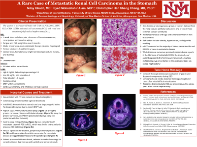
Introduction: RCC denotes a heterogeneous group of cancers derived from the renal tubular epithelial cells. While many types exist, Clear Cell RCC (ccRCC) constitutes most cases. In this report, we describe a case of ccRCC presenting ~10 years after radical nephrectomy as multiple gastric polyps.
Case Description/Methods: An 82-year-old male was evaluated with a 1-week history of chest pain, SOB, constipation, blood with wiping, and a 25lb unintentional weight loss over the past 6 months. He denied hematemesis, BRBPR, history of PUD, diverticulosis, or IBD. Relevant medical history included CAD s/p PCI in 2005, RCC with renal vein invasion s/p left radical nephrectomy in 2013, and GERD. The patient reported taking omeprazole as well as being on DAPT (held on admission) with aspirin and clopidogrel since his PCI as a “personal choice”. He had a strong smoking history and physical exam was unremarkable.
Admission labs did not reveal coagulopathy and the BMP was normal. CBC showed severe anemia (Hgb 6.3 g/dL), low iron of 16 µg/dL, low iron saturation of 4%, and hyperbilirubinemia (total bilirubin 1.5 mg/dL). All cardiac, pulmonary, and infectious workup was negative. In the ED the patient received one unit of packed RBCs, and recheck Hgb was 6.9.
Colonoscopy was significant for small-mouthed sigmoid diverticula, but no definitive cause for bleeding and subsequent anemia. EGD noted a 30mm and 40mm pedunculated polyp along the posterior wall of the cardia, both of which were removed using hot snare. Pathology of both polyps returned with morphology consistent with metastatic ccRCC.
Subsequent imaging included a PET/CT, which showed bilateral, peripheral predominant pulmonary lesions along with mediastinal and hilar lymphadenopathy. Following tumor board evaluation, the patient was referred to medical oncology for consideration of dual therapy with axitinib and pembrolizumab.
Discussion: There have been several reports in the literature noting gastric metastases from RCC. However, these have overwhelmingly – 1) involved patients with single metastases and 2) have occurred in patients who did not undergo radical nephrectomy. Our case is unique in that it describes the first known instance in the literature of a multiple metastatic polyp presentation in the cardia and body. Furthermore, it illustrates the importance of a thorough endoscopic evaluation, especially in cases of an initial difficult visualization. Clinicians must not be deterred from repeating EGD in such cases, as potential pathologies may be missed.

Disclosures:
Niloy Ghosh, MD, Syed Alam, MD, Christopher Chang, MD, PhD. P3877 - A Rare Case of Metastatic Renal Cell Carcinoma in the Stomach, ACG 2023 Annual Scientific Meeting Abstracts. Vancouver, BC, Canada: American College of Gastroenterology.
University of New Mexico Health Sciences Center, Albuquerque, NM
Introduction: RCC denotes a heterogeneous group of cancers derived from the renal tubular epithelial cells. While many types exist, Clear Cell RCC (ccRCC) constitutes most cases. In this report, we describe a case of ccRCC presenting ~10 years after radical nephrectomy as multiple gastric polyps.
Case Description/Methods: An 82-year-old male was evaluated with a 1-week history of chest pain, SOB, constipation, blood with wiping, and a 25lb unintentional weight loss over the past 6 months. He denied hematemesis, BRBPR, history of PUD, diverticulosis, or IBD. Relevant medical history included CAD s/p PCI in 2005, RCC with renal vein invasion s/p left radical nephrectomy in 2013, and GERD. The patient reported taking omeprazole as well as being on DAPT (held on admission) with aspirin and clopidogrel since his PCI as a “personal choice”. He had a strong smoking history and physical exam was unremarkable.
Admission labs did not reveal coagulopathy and the BMP was normal. CBC showed severe anemia (Hgb 6.3 g/dL), low iron of 16 µg/dL, low iron saturation of 4%, and hyperbilirubinemia (total bilirubin 1.5 mg/dL). All cardiac, pulmonary, and infectious workup was negative. In the ED the patient received one unit of packed RBCs, and recheck Hgb was 6.9.
Colonoscopy was significant for small-mouthed sigmoid diverticula, but no definitive cause for bleeding and subsequent anemia. EGD noted a 30mm and 40mm pedunculated polyp along the posterior wall of the cardia, both of which were removed using hot snare. Pathology of both polyps returned with morphology consistent with metastatic ccRCC.
Subsequent imaging included a PET/CT, which showed bilateral, peripheral predominant pulmonary lesions along with mediastinal and hilar lymphadenopathy. Following tumor board evaluation, the patient was referred to medical oncology for consideration of dual therapy with axitinib and pembrolizumab.
Discussion: There have been several reports in the literature noting gastric metastases from RCC. However, these have overwhelmingly – 1) involved patients with single metastases and 2) have occurred in patients who did not undergo radical nephrectomy. Our case is unique in that it describes the first known instance in the literature of a multiple metastatic polyp presentation in the cardia and body. Furthermore, it illustrates the importance of a thorough endoscopic evaluation, especially in cases of an initial difficult visualization. Clinicians must not be deterred from repeating EGD in such cases, as potential pathologies may be missed.

Figure: Metastatic RCC in the Stomach
a) 30mm Pedunculated Polyp
b) 10mm multilobulated polyp
c) Gastric polyp histopathology
d) Prior RCC histopathology
e-g) PET/CT evidence of metastasis
a) 30mm Pedunculated Polyp
b) 10mm multilobulated polyp
c) Gastric polyp histopathology
d) Prior RCC histopathology
e-g) PET/CT evidence of metastasis
Disclosures:
Niloy Ghosh indicated no relevant financial relationships.
Syed Alam indicated no relevant financial relationships.
Christopher Chang indicated no relevant financial relationships.
Niloy Ghosh, MD, Syed Alam, MD, Christopher Chang, MD, PhD. P3877 - A Rare Case of Metastatic Renal Cell Carcinoma in the Stomach, ACG 2023 Annual Scientific Meeting Abstracts. Vancouver, BC, Canada: American College of Gastroenterology.
