Tuesday Poster Session
Category: Liver
P3912 - Gallbladder Metastasis as an Atypical Presentation of Hepatocellular Carcinoma
Tuesday, October 24, 2023
10:30 AM - 4:00 PM PT
Location: Exhibit Hall

Has Audio

Zahra Dossaji, DO
Kirk Kerkorian School of Medicine at UNLV
Las Vegas, NV
Presenting Author(s)
Zahra Dossaji, DO1, Rajan Amin, MD2, Tooba Laeeq, MD3, Mark Hsu, MD1, Adam Khattak, DO1, Shahid Wahid, MD1
1Kirk Kerkorian School of Medicine at UNLV, Las Vegas, NV; 2University of Nevada, Las Vegas, Las Vegas, NV; 3University of Nevada, Las Vegas, Henderson, NV
Introduction: Hepatocellular carcinoma (HCC) is the third leading cause of cancer-related deaths worldwide, with an annual incidence of 2-6% in cirrhotic patients. The most common sites of metastasis are the lungs, lymph nodes, and bones. HCC metastasis to the gallbladder is rare due to HCC's infrequent invasion of the muscle layer and collagen fibers of the gallbladder wall. Here we present a case of HCC with metastasis to the gallbladder.
Case Description/Methods: A 73-year-old man with a history of hypertension and diabetes presented with a 2-month history of nausea, vomiting, diarrhea, chills, and right upper quadrant pain. He denied any history of alcohol or illicit drug use, recent travel, or sick contacts. He reported a family history of colon and gastric cancer.
Laboratory studies revealed an AFP of 39, WBC 12, ALT 45, AST 46, Total bilirubin 4.2, and Alk Phos 228. Hepatitis serologies and HIV were negative. CT abdomen revealed a new diagnosis of cirrhosis with chronic thrombosis of the portal vein and numerous small hepatic hypodense lesions. A follow-up MRI liver protocol was concerning for carcinoma. Endoscopic ultrasound revealed multiple hepatic lesions which were not amenable to biopsy.
Four months later, the patient underwent elective cholecystectomy. Final pathology of the gallbladder revealed several tumor aggregates within the gallbladder wall and mostly within vascular structures. Immunohistochemical tumor nodules stained positively for keratin CAM 5.2, arginase, and Heppar-1, consistent with metastatic hepatocellular carcinoma to the gallbladder (Figure 1). The patient was referred to medical oncology for further management.
Discussion: Metastatic HCC to the gallbladder is very rare, with an autopsy case series of 1000 patients noting the incidence to be 5.8%. The most used and recommended imaging methods are multiphasic CT and MRI. Physicians should maintain a high level of clinical suspicion as early detection is crucial for the early diagnosis and management of HCC. HCC metastasis to the gallbladder should be included in the differential diagnosis, despite its rarity. Our aim is to increase awareness, as prompt identification and timely intervention can improve patient outcomes.

Disclosures:
Zahra Dossaji, DO1, Rajan Amin, MD2, Tooba Laeeq, MD3, Mark Hsu, MD1, Adam Khattak, DO1, Shahid Wahid, MD1. P3912 - Gallbladder Metastasis as an Atypical Presentation of Hepatocellular Carcinoma, ACG 2023 Annual Scientific Meeting Abstracts. Vancouver, BC, Canada: American College of Gastroenterology.
1Kirk Kerkorian School of Medicine at UNLV, Las Vegas, NV; 2University of Nevada, Las Vegas, Las Vegas, NV; 3University of Nevada, Las Vegas, Henderson, NV
Introduction: Hepatocellular carcinoma (HCC) is the third leading cause of cancer-related deaths worldwide, with an annual incidence of 2-6% in cirrhotic patients. The most common sites of metastasis are the lungs, lymph nodes, and bones. HCC metastasis to the gallbladder is rare due to HCC's infrequent invasion of the muscle layer and collagen fibers of the gallbladder wall. Here we present a case of HCC with metastasis to the gallbladder.
Case Description/Methods: A 73-year-old man with a history of hypertension and diabetes presented with a 2-month history of nausea, vomiting, diarrhea, chills, and right upper quadrant pain. He denied any history of alcohol or illicit drug use, recent travel, or sick contacts. He reported a family history of colon and gastric cancer.
Laboratory studies revealed an AFP of 39, WBC 12, ALT 45, AST 46, Total bilirubin 4.2, and Alk Phos 228. Hepatitis serologies and HIV were negative. CT abdomen revealed a new diagnosis of cirrhosis with chronic thrombosis of the portal vein and numerous small hepatic hypodense lesions. A follow-up MRI liver protocol was concerning for carcinoma. Endoscopic ultrasound revealed multiple hepatic lesions which were not amenable to biopsy.
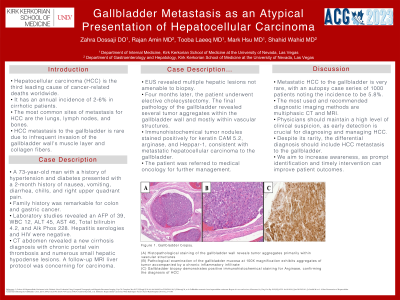
Four months later, the patient underwent elective cholecystectomy. Final pathology of the gallbladder revealed several tumor aggregates within the gallbladder wall and mostly within vascular structures. Immunohistochemical tumor nodules stained positively for keratin CAM 5.2, arginase, and Heppar-1, consistent with metastatic hepatocellular carcinoma to the gallbladder (Figure 1). The patient was referred to medical oncology for further management.
Discussion: Metastatic HCC to the gallbladder is very rare, with an autopsy case series of 1000 patients noting the incidence to be 5.8%. The most used and recommended imaging methods are multiphasic CT and MRI. Physicians should maintain a high level of clinical suspicion as early detection is crucial for the early diagnosis and management of HCC. HCC metastasis to the gallbladder should be included in the differential diagnosis, despite its rarity. Our aim is to increase awareness, as prompt identification and timely intervention can improve patient outcomes.

Figure: Figure 1. Gallbladder biopsy.
(A) Histopathological staining of the gallbladder wall reveals aggregates of tumor primarily within vascular structures (hematoxylin and eosin, 40x magnification).
(B) Pathological examination of the gallbladder mucosa exhibits aggregates of tumor accompanied by a chronic inflammatory infiltrate (hematoxylin and eosin, 100x magnification).
(C) Gallbladder biopsy demonstrates positive immunohistochemical staining for Arginase, confirming the diagnosis of HCC (100x magnification).
Disclosures:
Zahra Dossaji indicated no relevant financial relationships.
Rajan Amin indicated no relevant financial relationships.
Tooba Laeeq indicated no relevant financial relationships.
Mark Hsu indicated no relevant financial relationships.
Adam Khattak indicated no relevant financial relationships.
Shahid Wahid indicated no relevant financial relationships.
Zahra Dossaji, DO1, Rajan Amin, MD2, Tooba Laeeq, MD3, Mark Hsu, MD1, Adam Khattak, DO1, Shahid Wahid, MD1. P3912 - Gallbladder Metastasis as an Atypical Presentation of Hepatocellular Carcinoma, ACG 2023 Annual Scientific Meeting Abstracts. Vancouver, BC, Canada: American College of Gastroenterology.
