Tuesday Poster Session
Category: Biliary/Pancreas
P2913 - Boulder Dash: Endoscopic Management of Bouveret Syndrome and Gallstone Ileus
Tuesday, October 24, 2023
10:30 AM - 4:00 PM PT
Location: Exhibit Hall

Has Audio
- WH
William Hirsch, MD
University of Minnesota
Minneapolis, MN
Presenting Author(s)
William Hirsch, MD, Bryant Megna, MD, Nabeel Azeem, MD
University of Minnesota, Minneapolis, MN
Introduction: Gallstone ileus and Bouveret Syndrome are extraordinarily rare complications of gallstone disease. Given the scarcity of available data, little is known about best practices for medical, surgical, and/or endoscopic management. Herein we describe the co-occurrence of Bouveret Syndrome with gallstone ileus and successful endoscopic management.
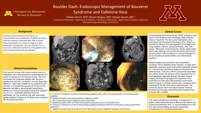
Case Description/Methods: A 68 year old woman with a past medical history of metastatic colon adenocarcinoma presented with two weeks of nausea and non-bloody emesis. She was tachycardic but otherwise afebrile with non-toxic appearance and soft, non-tender, and non-distended abdomen. Labs were notable for low potassium and chloride. A computed tomography (CT) scan of the abdomen and pelvis demonstrated pneumobilia related to a cholecystoduodenal fistula with migration of a 3.0 x 5.1 cm gallstone from the gallbladder into the duodenal bulb with associated gastric distention. Findings were consistent with Bouveret syndrome. An esophagogastroduodenoscopy (EGD) showed a large stone in the duodenal bulb completely obstructing the pylorus. The stone was fragmented using endoscopic electrohydraulic lithotripsy (EHL) followed by retrieval and dislodgement of the fragments using baskets, balloons, grasping forceps, nets, and snares. Afterwards, three temporary double pigtail plastic stents were deployed across the duodenal bulb into the second portion of the duodenum to ensure drainage as the surrounding duodenal edema and inflammation resolved. Repeat imaging was performed due to persistent symptoms which identified distal migration of large stone fragments causing a distal small bowel obstruction. Colonoscopy identified a malignant ileocecal (IC) valve stricture. A 25 mm x 90 mm uncovered metal stent was placed across the stricture with subsequent flow of small gallstone fragments through the stent. Repeat imaging illustrated large gallstone fragments in the proximal end of the stent with upstream small bowel dilation. Repeat colonoscopy visualized stone fragments in the left colon which were removed. The previously placed stent had fully expanded allowing traversal and the large obstructing stone fragment was removed with a basket.
Discussion: This case illustrates the endoscopic management of gastric outlet obstruction due to Bouveret syndrome and subsequent gallstone ileus. To our knowledge, this is the first case demonstrating successful endoscopic management of a combined presentation.

Disclosures:
William Hirsch, MD, Bryant Megna, MD, Nabeel Azeem, MD. P2913 - Boulder Dash: Endoscopic Management of Bouveret Syndrome and Gallstone Ileus, ACG 2023 Annual Scientific Meeting Abstracts. Vancouver, BC, Canada: American College of Gastroenterology.
University of Minnesota, Minneapolis, MN
Introduction: Gallstone ileus and Bouveret Syndrome are extraordinarily rare complications of gallstone disease. Given the scarcity of available data, little is known about best practices for medical, surgical, and/or endoscopic management. Herein we describe the co-occurrence of Bouveret Syndrome with gallstone ileus and successful endoscopic management.
Case Description/Methods: A 68 year old woman with a past medical history of metastatic colon adenocarcinoma presented with two weeks of nausea and non-bloody emesis. She was tachycardic but otherwise afebrile with non-toxic appearance and soft, non-tender, and non-distended abdomen. Labs were notable for low potassium and chloride. A computed tomography (CT) scan of the abdomen and pelvis demonstrated pneumobilia related to a cholecystoduodenal fistula with migration of a 3.0 x 5.1 cm gallstone from the gallbladder into the duodenal bulb with associated gastric distention. Findings were consistent with Bouveret syndrome. An esophagogastroduodenoscopy (EGD) showed a large stone in the duodenal bulb completely obstructing the pylorus. The stone was fragmented using endoscopic electrohydraulic lithotripsy (EHL) followed by retrieval and dislodgement of the fragments using baskets, balloons, grasping forceps, nets, and snares. Afterwards, three temporary double pigtail plastic stents were deployed across the duodenal bulb into the second portion of the duodenum to ensure drainage as the surrounding duodenal edema and inflammation resolved. Repeat imaging was performed due to persistent symptoms which identified distal migration of large stone fragments causing a distal small bowel obstruction. Colonoscopy identified a malignant ileocecal (IC) valve stricture. A 25 mm x 90 mm uncovered metal stent was placed across the stricture with subsequent flow of small gallstone fragments through the stent. Repeat imaging illustrated large gallstone fragments in the proximal end of the stent with upstream small bowel dilation. Repeat colonoscopy visualized stone fragments in the left colon which were removed. The previously placed stent had fully expanded allowing traversal and the large obstructing stone fragment was removed with a basket.
Discussion: This case illustrates the endoscopic management of gastric outlet obstruction due to Bouveret syndrome and subsequent gallstone ileus. To our knowledge, this is the first case demonstrating successful endoscopic management of a combined presentation.

Figure: A: Coronal CT abdomen and pelvis demonstrating gallstone within the duodenal bulb and resulting gastric distention
B: Gallstone located at the duodenal bulb visualized on EGD
C: EHL probe being used to fragment stone during EGD
D: Coronal CT abdomen and pelvis demonstrating gallstone fragment within the small bowel leading to small bowel obstruction
B: Gallstone located at the duodenal bulb visualized on EGD
C: EHL probe being used to fragment stone during EGD
D: Coronal CT abdomen and pelvis demonstrating gallstone fragment within the small bowel leading to small bowel obstruction
Disclosures:
William Hirsch indicated no relevant financial relationships.
Bryant Megna indicated no relevant financial relationships.
Nabeel Azeem: Boston Scientific – Consultant.
William Hirsch, MD, Bryant Megna, MD, Nabeel Azeem, MD. P2913 - Boulder Dash: Endoscopic Management of Bouveret Syndrome and Gallstone Ileus, ACG 2023 Annual Scientific Meeting Abstracts. Vancouver, BC, Canada: American College of Gastroenterology.
