Tuesday Poster Session
Category: Biliary/Pancreas
P2920 - Unveiling the Culprit: A Case of Acute Pancreatitis Unmasking Diffuse Large B-cell Lymphoma
Tuesday, October 24, 2023
10:30 AM - 4:00 PM PT
Location: Exhibit Hall

Has Audio

Tessa Herman, MD
University of Minnesota
Minneapolis, MN
Presenting Author(s)
Tessa Herman, MD1, Natalie Wilson, MD1, Mohamed Abdallah, MD2, Mohammad Bilal, MD3, Hashim Nemat, MBBS4
1University of Minnesota, Minneapolis, MN; 2University of Minnesota Medical Center, Minneapolis, MN; 3Minneapolis VA Medical Center, Minneapolis, MN; 4HealthPartners, Regions Hospital, St. Paul, MN
Introduction: Acute pancreatitis is a common cause of acute abdominal pain and can be due to a variety of causes, most often from gallstones or alcohol use. Malignancies such as lymphoma are infrequent causes of acute pancreatitis, especially as the initial presentation of malignancy. We present a case of acute pancreatitis caused by secondary pancreatic involvement of diffuse large B-cell lymphoma (DLBCL).
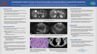
Case Description/Methods: A previously healthy 27-year-old male presented with acute epigastric pain and was found to have a serum lipase of 2,689 U/L, consistent with acute pancreatitis. Liver function tests were normal. Abdominal ultrasound showed a 4.1 cm hypoechoic cystic lesion in the pancreatic head without biliary obstruction. Follow-up MRI confirmed a 3.5 x 3.4 cm solid mass in the pancreatic tail [Figure 1A] and a 3.9 x 3.1 cm solid mass in the pancreatic head [Figure 1B]. An endoscopic ultrasound (EUS) was performed and fine needle biopsies from both the masses revealed DLBCL. The patient subsequently developed elevated liver function tests with imaging showing common bile duct stenosis secondary to the pancreatic head mass. Endoscopic retrograde cholangiopancreatography (ERCP) was attempted but the duodenoscope was unable to pass the upper esophagus despite dilation. The procedure was aborted after a longitudinal mucosal tear in this area was noted. CT scan of the chest revealed a large anterior mediastinal mass with extrinsic compression, later confirmed to be DLBCL [Figure 1C]. Given his abnormal liver function tests, he was not eligible for standard chemotherapy and was instead treated with bendamustine, rituximab, steroids, and a percutaneous biliary drain was placed for palliation of jaundice prior to initiation of standard chemotherapy.
Discussion: DLBCL commonly involves extranodal sites, including the gastrointestinal tract, although usually the involvement is limited to the stomach or small intestine. Acute pancreatitis can present as a first sign of malignancy, however, this presentation is rare in younger individuals. Our case highlights that malignancies such as lymphomas should be considered as a cause of acute pancreatitis in younger individuals when other common etiologies are excluded.

Disclosures:
Tessa Herman, MD1, Natalie Wilson, MD1, Mohamed Abdallah, MD2, Mohammad Bilal, MD3, Hashim Nemat, MBBS4. P2920 - Unveiling the Culprit: A Case of Acute Pancreatitis Unmasking Diffuse Large B-cell Lymphoma, ACG 2023 Annual Scientific Meeting Abstracts. Vancouver, BC, Canada: American College of Gastroenterology.
1University of Minnesota, Minneapolis, MN; 2University of Minnesota Medical Center, Minneapolis, MN; 3Minneapolis VA Medical Center, Minneapolis, MN; 4HealthPartners, Regions Hospital, St. Paul, MN
Introduction: Acute pancreatitis is a common cause of acute abdominal pain and can be due to a variety of causes, most often from gallstones or alcohol use. Malignancies such as lymphoma are infrequent causes of acute pancreatitis, especially as the initial presentation of malignancy. We present a case of acute pancreatitis caused by secondary pancreatic involvement of diffuse large B-cell lymphoma (DLBCL).
Case Description/Methods: A previously healthy 27-year-old male presented with acute epigastric pain and was found to have a serum lipase of 2,689 U/L, consistent with acute pancreatitis. Liver function tests were normal. Abdominal ultrasound showed a 4.1 cm hypoechoic cystic lesion in the pancreatic head without biliary obstruction. Follow-up MRI confirmed a 3.5 x 3.4 cm solid mass in the pancreatic tail [Figure 1A] and a 3.9 x 3.1 cm solid mass in the pancreatic head [Figure 1B]. An endoscopic ultrasound (EUS) was performed and fine needle biopsies from both the masses revealed DLBCL. The patient subsequently developed elevated liver function tests with imaging showing common bile duct stenosis secondary to the pancreatic head mass. Endoscopic retrograde cholangiopancreatography (ERCP) was attempted but the duodenoscope was unable to pass the upper esophagus despite dilation. The procedure was aborted after a longitudinal mucosal tear in this area was noted. CT scan of the chest revealed a large anterior mediastinal mass with extrinsic compression, later confirmed to be DLBCL [Figure 1C]. Given his abnormal liver function tests, he was not eligible for standard chemotherapy and was instead treated with bendamustine, rituximab, steroids, and a percutaneous biliary drain was placed for palliation of jaundice prior to initiation of standard chemotherapy.
Discussion: DLBCL commonly involves extranodal sites, including the gastrointestinal tract, although usually the involvement is limited to the stomach or small intestine. Acute pancreatitis can present as a first sign of malignancy, however, this presentation is rare in younger individuals. Our case highlights that malignancies such as lymphomas should be considered as a cause of acute pancreatitis in younger individuals when other common etiologies are excluded.

Figure: Figure 1 - Radiographic imaging of the pancreatic and mediastinal masses. MRI images (T2 FLAIR sequence) reveal a 3.5 x 3.4 cm solid mass in the pancreatic tail (1A) and a 3.9 x 3.1 cm solid mass in the pancreatic head (1B). CT chest with contrast reveals a large anterior mediastinal mass that encases the origin of the great vessels and compresses the trachea (greater than 50% narrowing just above the carina) (1C).
Disclosures:
Tessa Herman indicated no relevant financial relationships.
Natalie Wilson indicated no relevant financial relationships.
Mohamed Abdallah indicated no relevant financial relationships.
Mohammad Bilal: Boston Scientific – Consultant.
Hashim Nemat indicated no relevant financial relationships.
Tessa Herman, MD1, Natalie Wilson, MD1, Mohamed Abdallah, MD2, Mohammad Bilal, MD3, Hashim Nemat, MBBS4. P2920 - Unveiling the Culprit: A Case of Acute Pancreatitis Unmasking Diffuse Large B-cell Lymphoma, ACG 2023 Annual Scientific Meeting Abstracts. Vancouver, BC, Canada: American College of Gastroenterology.
