Tuesday Poster Session
Category: Biliary/Pancreas
P2936 - Hairy Cell Leukemia: A Rare Differential for a Pancreatic Mass
Tuesday, October 24, 2023
10:30 AM - 4:00 PM PT
Location: Exhibit Hall

Has Audio
.jpg)
Nishant Aggarwal, MD
William Beaumont Hospital-Royal Oak
Royal Oak, MI
Presenting Author(s)
Nishant Aggarwal, MD, Rabin Neupane, MD, Christopher Thorburn, MD, Mohammad M. Chisti, MD, Laith H. Jamil, MD, FACG
William Beaumont Hospital-Royal Oak, Royal Oak, MI
Introduction: Hairy cell leukemia (HCL) is a B-cell lymphoproliferative disorder characterized by the presence of neoplastic B-cells with cytoplasmic villi (hairy) mainly in peripheral blood, bone marrow, and splenic red pulp, and sometimes also in liver, lymph nodes, or bones. Here, we present a patient with a rare case of HCL presenting as a pancreatic mass.
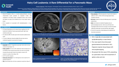
Case Description/Methods: A 62-year-old gentleman presented with right lower extremity sciatic pain and underwent magnetic resonance imaging (MRI) of lumbar spine which showed incidental renal mass. His medical history was significant for HCL, treated with cladribine 9 years ago. Computed tomography of abdomen showed multiple left renal cysts and an incidental irregularity in pancreatic tail margins. Initial MRI abdomen confirmed the presence of irregularity at pancreatic tail but did not show a discrete mass. Follow-up MRI after nine months showed significant interval progression of an infiltrative lesion along the pancreatic tail, which was partially encasing the pancreatic body, tail and splenic vessels, with extension to splenic hilum (figure 1A). CA 19-9 and CEA were negative. Endoscopic ultrasound showed a 6.3 cm heterogenous solid mass suspicious of lymphoma (figure 1B). Fine-needle aspiration revealed small lymphocytes with hairy cytoplasmic projections (figure 1C) and flow cytometry confirmed expression of pan-B cell markers in addition to CD11c, CD25, CD103 and kappa light chains, consistent with HCL. Positron emission tomography scan revealed an FDG-avid infiltrating lesion in pancreatic body and tail. The esophagogastroduodenoscopy also showed an incidental fungating mass at gastroesophageal junction, which was confirmed to be esophageal adenocarcinoma on biopsy, for which the patient underwent endoscopic mucosal resection after staging. For his HCL recurrence, chemotherapy with cladribine and rituximab was planned.
Discussion: HCL is typically not associated with lymphadenopathy or mass lesions. Few case reports have discussed gastrointestinal involvement in HCL. Diagnosis requires tissue biopsy with immunophenotyping. We present the first case of HCL presenting as a pancreatic mass with encasement of splenic artery and vein.

Disclosures:
Nishant Aggarwal, MD, Rabin Neupane, MD, Christopher Thorburn, MD, Mohammad M. Chisti, MD, Laith H. Jamil, MD, FACG. P2936 - Hairy Cell Leukemia: A Rare Differential for a Pancreatic Mass, ACG 2023 Annual Scientific Meeting Abstracts. Vancouver, BC, Canada: American College of Gastroenterology.
William Beaumont Hospital-Royal Oak, Royal Oak, MI
Introduction: Hairy cell leukemia (HCL) is a B-cell lymphoproliferative disorder characterized by the presence of neoplastic B-cells with cytoplasmic villi (hairy) mainly in peripheral blood, bone marrow, and splenic red pulp, and sometimes also in liver, lymph nodes, or bones. Here, we present a patient with a rare case of HCL presenting as a pancreatic mass.
Case Description/Methods: A 62-year-old gentleman presented with right lower extremity sciatic pain and underwent magnetic resonance imaging (MRI) of lumbar spine which showed incidental renal mass. His medical history was significant for HCL, treated with cladribine 9 years ago. Computed tomography of abdomen showed multiple left renal cysts and an incidental irregularity in pancreatic tail margins. Initial MRI abdomen confirmed the presence of irregularity at pancreatic tail but did not show a discrete mass. Follow-up MRI after nine months showed significant interval progression of an infiltrative lesion along the pancreatic tail, which was partially encasing the pancreatic body, tail and splenic vessels, with extension to splenic hilum (figure 1A). CA 19-9 and CEA were negative. Endoscopic ultrasound showed a 6.3 cm heterogenous solid mass suspicious of lymphoma (figure 1B). Fine-needle aspiration revealed small lymphocytes with hairy cytoplasmic projections (figure 1C) and flow cytometry confirmed expression of pan-B cell markers in addition to CD11c, CD25, CD103 and kappa light chains, consistent with HCL. Positron emission tomography scan revealed an FDG-avid infiltrating lesion in pancreatic body and tail. The esophagogastroduodenoscopy also showed an incidental fungating mass at gastroesophageal junction, which was confirmed to be esophageal adenocarcinoma on biopsy, for which the patient underwent endoscopic mucosal resection after staging. For his HCL recurrence, chemotherapy with cladribine and rituximab was planned.
Discussion: HCL is typically not associated with lymphadenopathy or mass lesions. Few case reports have discussed gastrointestinal involvement in HCL. Diagnosis requires tissue biopsy with immunophenotyping. We present the first case of HCL presenting as a pancreatic mass with encasement of splenic artery and vein.

Figure: Figure 1: (A) Magnetic resonance imaging scan of pancreas showing a 6.5 x 6 cm infiltrative lesion extending along the pancreatic tail, with encasement of splenic vessels and hilum, (B) Endoscopic ultrasound showing a 6.3 x 4.2 cm irregular mass in the pancreatic tail for which fine needle aspiration was performed. (C) Pancreatic mass cytology showing small lymphocytes (low power view) and hairy cytoplasmic projections (inset; high power view) suggestive of hairy cell leukemia.
Disclosures:
Nishant Aggarwal indicated no relevant financial relationships.
Rabin Neupane indicated no relevant financial relationships.
Christopher Thorburn indicated no relevant financial relationships.
Mohammad Chisti indicated no relevant financial relationships.
Laith Jamil indicated no relevant financial relationships.
Nishant Aggarwal, MD, Rabin Neupane, MD, Christopher Thorburn, MD, Mohammad M. Chisti, MD, Laith H. Jamil, MD, FACG. P2936 - Hairy Cell Leukemia: A Rare Differential for a Pancreatic Mass, ACG 2023 Annual Scientific Meeting Abstracts. Vancouver, BC, Canada: American College of Gastroenterology.
