Tuesday Poster Session
Category: Biliary/Pancreas
P2944 - A Rare Case of Malignant Metastatic Melanoma to the Pancreas
Tuesday, October 24, 2023
10:30 AM - 4:00 PM PT
Location: Exhibit Hall

Has Audio
- RA
Raya Al-Ashram, MD
Albany Medical Center
Albany, NY
Presenting Author(s)
Ahmad Abulawi, MD1, Raya Alashram, MD2, Omar Tageldin, MBBCh1, Stephen Hasak, MD, MPH1
1Albany Medical College, Albany, NY; 2Albany Medical Center, Albany, NY
Introduction: The most common primary malignancies that metastasize to the pancreas include kidney, lung, colon, and breast cancers, however, the metastatic disease of the pancreas is rare and accounts for 2 – 5 % of the pancreatic malignancies. Metastatic melanoma in the pancreas is one of the unusual locations with very few reported cases in the literature.
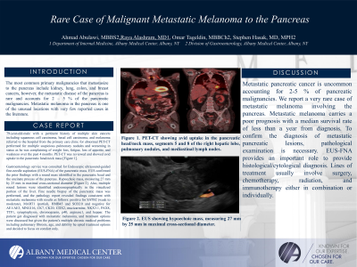
Case Description/Methods: 79-year-old-male with a pertinent history of multiple skin cancers including squamous cell carcinoma, basal cell carcinoma, and melanoma referred to the hospital from the primary care clinic for abnormal PET-CT performed for multiple suspicious pulmonary nodules and worsening in status as he was complaining of weight loss, fatigue, loss of appetite, and weakness over the past 4 months. PET-CT was reviewed and showed avid uptake in the pancreatic head/neck mass [Figure 1].
Gastroenterology service was consulted for Endoscopic ultrasound-guided fine-needle aspiration (EUS-FNA) of the pancreatic mass. EUS confirmed the prior findings with a round mass identified in the pancreatic head and the uncinate process of the pancreas. Hypoechoic mass, measuring 27 mm by 25 mm in maximal cross-sectional diameter [Figure 2]. Also, multiple round lesions were identified endosonographically in the visualized portion of the liver. Fine needle biopsy of the pancreatic mass was performed, and the pathology report revealed findings consistent with metastatic melanoma with results as follows: positive for SATB2 (weak to moderate), MART1 (partial), HMB45 and SOX10 and negative for AE1/AE3, MNG116, CK7, CK20, CDX2, mucicarmine, NKX3.1, PAX8, TTF1, synaptophysin, chromogranin, p40, arginase1, and heppar. The patient got diagnosed with metastatic melanoma, and treatment options were discussed but given the patient’s multiple chronic medical problems including pulmonary fibrosis, age, and debility he opted treatment options and decided to focus on comfort only.
Discussion: Metastatic pancreatic cancer is uncommon accounting for 2-5 % of pancreatic malignancies. We report a very rare case of metastatic melanoma involving the pancreas. Metastatic melanoma carries a poor prognosis with a median survival rate of less than a year from diagnosis. To confirm the diagnosis of metastatic pancreatic lesions, pathological examination is necessary. EUS-FNA provides an important role to provide histological/cytological diagnosis. Lines of treatment usually involve surgery, chemotherapy, radiation, and immunotherapy either in combination or individually.

Disclosures:
Ahmad Abulawi, MD1, Raya Alashram, MD2, Omar Tageldin, MBBCh1, Stephen Hasak, MD, MPH1. P2944 - A Rare Case of Malignant Metastatic Melanoma to the Pancreas, ACG 2023 Annual Scientific Meeting Abstracts. Vancouver, BC, Canada: American College of Gastroenterology.
1Albany Medical College, Albany, NY; 2Albany Medical Center, Albany, NY
Introduction: The most common primary malignancies that metastasize to the pancreas include kidney, lung, colon, and breast cancers, however, the metastatic disease of the pancreas is rare and accounts for 2 – 5 % of the pancreatic malignancies. Metastatic melanoma in the pancreas is one of the unusual locations with very few reported cases in the literature.
Case Description/Methods: 79-year-old-male with a pertinent history of multiple skin cancers including squamous cell carcinoma, basal cell carcinoma, and melanoma referred to the hospital from the primary care clinic for abnormal PET-CT performed for multiple suspicious pulmonary nodules and worsening in status as he was complaining of weight loss, fatigue, loss of appetite, and weakness over the past 4 months. PET-CT was reviewed and showed avid uptake in the pancreatic head/neck mass [Figure 1].
Gastroenterology service was consulted for Endoscopic ultrasound-guided fine-needle aspiration (EUS-FNA) of the pancreatic mass. EUS confirmed the prior findings with a round mass identified in the pancreatic head and the uncinate process of the pancreas. Hypoechoic mass, measuring 27 mm by 25 mm in maximal cross-sectional diameter [Figure 2]. Also, multiple round lesions were identified endosonographically in the visualized portion of the liver. Fine needle biopsy of the pancreatic mass was performed, and the pathology report revealed findings consistent with metastatic melanoma with results as follows: positive for SATB2 (weak to moderate), MART1 (partial), HMB45 and SOX10 and negative for AE1/AE3, MNG116, CK7, CK20, CDX2, mucicarmine, NKX3.1, PAX8, TTF1, synaptophysin, chromogranin, p40, arginase1, and heppar. The patient got diagnosed with metastatic melanoma, and treatment options were discussed but given the patient’s multiple chronic medical problems including pulmonary fibrosis, age, and debility he opted treatment options and decided to focus on comfort only.
Discussion: Metastatic pancreatic cancer is uncommon accounting for 2-5 % of pancreatic malignancies. We report a very rare case of metastatic melanoma involving the pancreas. Metastatic melanoma carries a poor prognosis with a median survival rate of less than a year from diagnosis. To confirm the diagnosis of metastatic pancreatic lesions, pathological examination is necessary. EUS-FNA provides an important role to provide histological/cytological diagnosis. Lines of treatment usually involve surgery, chemotherapy, radiation, and immunotherapy either in combination or individually.

Figure: Figure 1: PET-CT showing avid uptake in the pancreatic head/neck mass, segments 5 and 8 of the right hepatic lobe, pulmonary nodules, and mediastinal lymph nodes.
Figure 2: EUS showing hypoechoic mass, measuring 27 mm by 25 mm in maximal cross-sectional diameter.
Figure 2: EUS showing hypoechoic mass, measuring 27 mm by 25 mm in maximal cross-sectional diameter.
Disclosures:
Ahmad Abulawi indicated no relevant financial relationships.
Raya Alashram indicated no relevant financial relationships.
Omar Tageldin indicated no relevant financial relationships.
Stephen Hasak indicated no relevant financial relationships.
Ahmad Abulawi, MD1, Raya Alashram, MD2, Omar Tageldin, MBBCh1, Stephen Hasak, MD, MPH1. P2944 - A Rare Case of Malignant Metastatic Melanoma to the Pancreas, ACG 2023 Annual Scientific Meeting Abstracts. Vancouver, BC, Canada: American College of Gastroenterology.
