Tuesday Poster Session
Category: Liver
P3946 - Benign to Malignant Hepatic Lesion Transformation in Abernethy Malformation
Tuesday, October 24, 2023
10:30 AM - 4:00 PM PT
Location: Exhibit Hall

Has Audio

Chloe K. Tom, MD
Keck School of Medicine of USC
Los Angeles, CA
Presenting Author(s)
Chloe K. Tom, MD1, Ramon Ter-Oganesyan, MD2, Chopra Shefali, MD1, Navpreet Kaur, MD1, Jordan L. Pace, BS3, Ali Rastegarpour, MD1, Yuri Genyk, MD1, Jeffrey A. Kahn, MD4
1Keck School of Medicine of USC, Los Angeles, CA; 2University of Southern California, Los Angeles, CA; 3California University of Science and Medicine, Colton, CA; 4Keck School of Medicine, University of Southern California, Los Angeles, CA
Introduction: Abernethy malformation or congenital extrahepatic portosystemic shunt (CEPS) is an extremely rare condition whereby the porto-mesenteric blood drains into a systemic vein, bypassing the liver through a complete or partial shunt. Complications of CEPS include hyperammonemia, encephalopathy, benign and malignant liver tumors, and hepatopulmonary syndrome. We describe a case of an adult female with CEPS, who developed focal nodular hyperplasia (FNH) with subsequent malignant transformation into hepatocellular carcinoma (HCC).
Case Description/Methods: An adolescent female patient with precocious puberty underwent an ultrasound of the abdomen and pelvis as part of the workup for elevated sex hormones and was found to have multiple hepatic lesions. One lesion was biopsied, demonstrating FNH. Several years later at age 27, the patient presented to the hospital with pruritus. She had a normal body mass index, normal vital signs, and a physical exam that was notable for non-tender palpable hepatomegaly. Laboratory tests were notable for alkaline phosphatase 329 units/L, aspartate aminotransferase 72 units/L, alanine aminotransferase 85 units/L, and ferritin 200 ug/L. Total bilirubin, carcinoembryonic antigen, alpha-fetoprotein (AFP), and cancer antigen 19-9 were normal. Contrast-enhanced computed tomography (CT) showed multiple hepatic masses with disparate characteristics and hepatic vascular anatomy consistent with CEPS (Fig. A-C). One of the lesions was biopsied and demonstrated foci of HCC within a hepatic adenoma (Fig. D). Given the large burden of lesions and biopsy-proven HCC, the patient underwent living-donor liver transplantation. Microscopic evaluation of the explant demonstrated a lack of portal veins (Fig. E) and distinct types of lesions which consisted of 2 FNH, 2 simple hepatic adenomas, and 3 HCC arising in adenoma. Following liver transplantation, the patient has excellent functional status with normal allograft function and no evidence of HCC recurrence (Fig. F).
Discussion: This case is an unusual presentation of complications of CEPS. We propose that serial diagnostic imaging should be performed to monitor enlarging or atypical hepatic lesions in patients with Abernathy malformation. Suspicious lesions should be biopsied regardless of AFP levels since this marker is often normal even in the setting of HCC, as was the case in this patient. Liver transplantation is a viable treatment option for patients with symptomatic enlarging masses.

Disclosures:
Chloe K. Tom, MD1, Ramon Ter-Oganesyan, MD2, Chopra Shefali, MD1, Navpreet Kaur, MD1, Jordan L. Pace, BS3, Ali Rastegarpour, MD1, Yuri Genyk, MD1, Jeffrey A. Kahn, MD4. P3946 - Benign to Malignant Hepatic Lesion Transformation in Abernethy Malformation, ACG 2023 Annual Scientific Meeting Abstracts. Vancouver, BC, Canada: American College of Gastroenterology.
1Keck School of Medicine of USC, Los Angeles, CA; 2University of Southern California, Los Angeles, CA; 3California University of Science and Medicine, Colton, CA; 4Keck School of Medicine, University of Southern California, Los Angeles, CA
Introduction: Abernethy malformation or congenital extrahepatic portosystemic shunt (CEPS) is an extremely rare condition whereby the porto-mesenteric blood drains into a systemic vein, bypassing the liver through a complete or partial shunt. Complications of CEPS include hyperammonemia, encephalopathy, benign and malignant liver tumors, and hepatopulmonary syndrome. We describe a case of an adult female with CEPS, who developed focal nodular hyperplasia (FNH) with subsequent malignant transformation into hepatocellular carcinoma (HCC).
Case Description/Methods: An adolescent female patient with precocious puberty underwent an ultrasound of the abdomen and pelvis as part of the workup for elevated sex hormones and was found to have multiple hepatic lesions. One lesion was biopsied, demonstrating FNH. Several years later at age 27, the patient presented to the hospital with pruritus. She had a normal body mass index, normal vital signs, and a physical exam that was notable for non-tender palpable hepatomegaly. Laboratory tests were notable for alkaline phosphatase 329 units/L, aspartate aminotransferase 72 units/L, alanine aminotransferase 85 units/L, and ferritin 200 ug/L. Total bilirubin, carcinoembryonic antigen, alpha-fetoprotein (AFP), and cancer antigen 19-9 were normal. Contrast-enhanced computed tomography (CT) showed multiple hepatic masses with disparate characteristics and hepatic vascular anatomy consistent with CEPS (Fig. A-C). One of the lesions was biopsied and demonstrated foci of HCC within a hepatic adenoma (Fig. D). Given the large burden of lesions and biopsy-proven HCC, the patient underwent living-donor liver transplantation. Microscopic evaluation of the explant demonstrated a lack of portal veins (Fig. E) and distinct types of lesions which consisted of 2 FNH, 2 simple hepatic adenomas, and 3 HCC arising in adenoma. Following liver transplantation, the patient has excellent functional status with normal allograft function and no evidence of HCC recurrence (Fig. F).
Discussion: This case is an unusual presentation of complications of CEPS. We propose that serial diagnostic imaging should be performed to monitor enlarging or atypical hepatic lesions in patients with Abernathy malformation. Suspicious lesions should be biopsied regardless of AFP levels since this marker is often normal even in the setting of HCC, as was the case in this patient. Liver transplantation is a viable treatment option for patients with symptomatic enlarging masses.

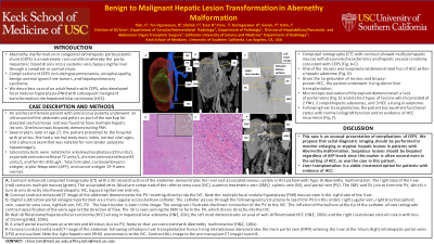
Figure: A: Contrast-enhanced computed tomography (CT) with a 3D reconstruction of the abdomen demonstrates the liver and associated venous system in this patient with Type Ib Abernethy malformation. The right lobe of the liver (red) contains multiple masses (green). The associated veins (blue) are comprised of the inferior vena cava (IVC), superior mesenteric vein (SMV), splenic vein (SV), and portal vein (PV). The SMV and SV join together to form the PV, which in turn drains directly into the extrahepatic IVC, bypassing the liver entirely.
B: Coronal contrast-enhanced CT image of the abdomen demonstrates the PV inserting directly into the IVC. Note the multiple focal nodular hyperplasia (FNH) masses seen in the right lobe of the liver.
C: Digital subtraction portal venogram performed via a trans-jugular access balloon catheter. The catheter passes through the following veins/structures to reach the PV (in this order): right jugular vein, right brachiocephalic vein, superior vena cava, right atrium, IVC, PV. The heart border is seen in the image. The venogram illustrates the direct connection of the PV to the IVC. The inflation of the balloon at the tip of the catheter allows retrograde contrast opacification of the veins against the direction of flow. The SV is seen joining the SMV to form the PV, which drains directly into the IVC.
D: Well-differentiated hepatocellular carcinoma (HCC) arising in hepatocellular adenoma (H&E, 20x); the left inset demonstrates an area of well-differentiated HCC (H&E, 200x) and the right inset shows reticulin stain with loss of staining (H&E, 200x).
E: A small portal tract shows an arteriole and bile duct, but no PV, features that are consistent with Abernethy malformation (H&E, 100x).
F: Coronal contrast-enhanced CT image of the abdomen following orthotopic liver transplantation from a living related donor demonstrates the main portal vein (MPV) entering the liver at the hilum. Right intrahepatic portal veins (IPV) are visualized. Note the right hepatic vein (RHV) anastomosis to the IVC. Contrast this image to the pre-transplant CT image in part B.
Disclosures:
Chloe Tom indicated no relevant financial relationships.
Ramon Ter-Oganesyan indicated no relevant financial relationships.
Chopra Shefali indicated no relevant financial relationships.
Navpreet Kaur indicated no relevant financial relationships.
Jordan Pace indicated no relevant financial relationships.
Ali Rastegarpour indicated no relevant financial relationships.
Yuri Genyk indicated no relevant financial relationships.
Jeffrey Kahn indicated no relevant financial relationships.
Chloe K. Tom, MD1, Ramon Ter-Oganesyan, MD2, Chopra Shefali, MD1, Navpreet Kaur, MD1, Jordan L. Pace, BS3, Ali Rastegarpour, MD1, Yuri Genyk, MD1, Jeffrey A. Kahn, MD4. P3946 - Benign to Malignant Hepatic Lesion Transformation in Abernethy Malformation, ACG 2023 Annual Scientific Meeting Abstracts. Vancouver, BC, Canada: American College of Gastroenterology.

