Tuesday Poster Session
Category: Biliary/Pancreas
P2948 - When the Pancreas Goes Rogue: A Tail of Aberrant Necrotizing Pancreatitis, and Gastroduodenal Pseudoaneurysm
Tuesday, October 24, 2023
10:30 AM - 4:00 PM PT
Location: Exhibit Hall

Has Audio

Afsheen Moshtaghi, DO
Midwestern University
Cottonwood, AZ
Presenting Author(s)
Afsheen Moshtaghi, DO1, Zachary M. Hanafi, DO2, Idrees Suliman, MD3, Rodney Engel, MD4
1Midwestern University, Cottonwood, AZ; 2Verde Valley Medical Center, Cottonwood, AZ; 3Midwestern University, Mesa, AZ; 4Flagstaff Medical Center, Flagstaff, AZ
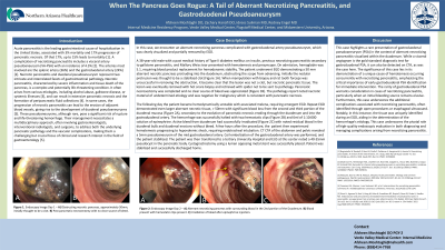
Introduction: Acute pancreatitis is the leading gastrointestinal cause of hospitalization in the United States, with 17% progression of pancreatic necrosis. 15% of which leads to mortality. One complication of necrotizing pancreatitis is visceral artery pseudoaneurysm (VA-PSA) with an incidence of 4.3%. The arteries involved include the splenic artery (36%) and the gastroduodenal artery (24%). In this case, we encounter an aberrant necrotizing pancreas complicated with gastroduodenal artery PSA, which was clearly seen and partially removed by non-invasive EGD.
Case Description/Methods: A 38-year-old male with a past medical history of gallstone pancreatitis presents with hematemesis and presyncope. On admission, hemoglobin was 5.1, requiring transfusion. The patient underwent EGD demonstrating a 50 mm aberrant necrotic pancreas in the duodenum. Attempted tissue confirmation was obtained using a snare. No bleeding was initially noted with the pathology report noted necrotic material. The following morning the patient became hypotensive with melena, requiring emergent EGD. Repeat EGD demonstrated even larger aberrant necrotic tissue > 50mm with significant bleeding in the second and third portions of the duodenum, likely secondary necrotic pancreas eroding through the duodenum. The bleed was halted with epinephrine and 2 hemostatic clips. The patient then experienced additional hematemesis, progressing to hypovolemic shock, requiring pressors. CTA of the abdomen and pelvis revealed a 5 mm pseudoaneurysm of the mid-gastroduodenal artery. Coil embolization was performed and the patient was stabilized.
Discussion: This case demonstrates a unique cause of hematemesis in the setting of necrotizing pancreatitis, as well as the importance of early identification of the gastroduodenal PSA for immediate intervention. A visceral angiogram is the gold standard diagnostic test, though it can be detected on CTA as in this case. Gastroduodenal PSA should be considered in necrotizing pancreatitis especially if a source of bleeding is not initially identified. Next, this case shows complications of necrotizing pancreatitis with aberrant pancreas which is usually identified by open procedures or esophageal ultrasound. This case identified the tissue on EGD which helped identify the etiology of the bleed. While a rare combination of issues, this case shows the importance of endoscopy skills in both the diagnosis and management of complications related to necrotizing pancreatitis.

Disclosures:
Afsheen Moshtaghi, DO1, Zachary M. Hanafi, DO2, Idrees Suliman, MD3, Rodney Engel, MD4. P2948 - When the Pancreas Goes Rogue: A Tail of Aberrant Necrotizing Pancreatitis, and Gastroduodenal Pseudoaneurysm, ACG 2023 Annual Scientific Meeting Abstracts. Vancouver, BC, Canada: American College of Gastroenterology.
1Midwestern University, Cottonwood, AZ; 2Verde Valley Medical Center, Cottonwood, AZ; 3Midwestern University, Mesa, AZ; 4Flagstaff Medical Center, Flagstaff, AZ
Introduction: Acute pancreatitis is the leading gastrointestinal cause of hospitalization in the United States, with 17% progression of pancreatic necrosis. 15% of which leads to mortality. One complication of necrotizing pancreatitis is visceral artery pseudoaneurysm (VA-PSA) with an incidence of 4.3%. The arteries involved include the splenic artery (36%) and the gastroduodenal artery (24%). In this case, we encounter an aberrant necrotizing pancreas complicated with gastroduodenal artery PSA, which was clearly seen and partially removed by non-invasive EGD.
Case Description/Methods: A 38-year-old male with a past medical history of gallstone pancreatitis presents with hematemesis and presyncope. On admission, hemoglobin was 5.1, requiring transfusion. The patient underwent EGD demonstrating a 50 mm aberrant necrotic pancreas in the duodenum. Attempted tissue confirmation was obtained using a snare. No bleeding was initially noted with the pathology report noted necrotic material. The following morning the patient became hypotensive with melena, requiring emergent EGD. Repeat EGD demonstrated even larger aberrant necrotic tissue > 50mm with significant bleeding in the second and third portions of the duodenum, likely secondary necrotic pancreas eroding through the duodenum. The bleed was halted with epinephrine and 2 hemostatic clips. The patient then experienced additional hematemesis, progressing to hypovolemic shock, requiring pressors. CTA of the abdomen and pelvis revealed a 5 mm pseudoaneurysm of the mid-gastroduodenal artery. Coil embolization was performed and the patient was stabilized.
Discussion: This case demonstrates a unique cause of hematemesis in the setting of necrotizing pancreatitis, as well as the importance of early identification of the gastroduodenal PSA for immediate intervention. A visceral angiogram is the gold standard diagnostic test, though it can be detected on CTA as in this case. Gastroduodenal PSA should be considered in necrotizing pancreatitis especially if a source of bleeding is not initially identified. Next, this case shows complications of necrotizing pancreatitis with aberrant pancreas which is usually identified by open procedures or esophageal ultrasound. This case identified the tissue on EGD which helped identify the etiology of the bleed. While a rare combination of issues, this case shows the importance of endoscopy skills in both the diagnosis and management of complications related to necrotizing pancreatitis.

Figure: Figure 1: Endoscopy Image
a: Aberrant necrotizing pancreas with surrounding blood in the 2nd portion of the Duodenum
b: Blood present with hemostatic clips present
c: Irradiation of bleed after epinephrine injection
a: Aberrant necrotizing pancreas with surrounding blood in the 2nd portion of the Duodenum
b: Blood present with hemostatic clips present
c: Irradiation of bleed after epinephrine injection
Disclosures:
Afsheen Moshtaghi indicated no relevant financial relationships.
Zachary Hanafi indicated no relevant financial relationships.
Idrees Suliman indicated no relevant financial relationships.
Rodney Engel indicated no relevant financial relationships.
Afsheen Moshtaghi, DO1, Zachary M. Hanafi, DO2, Idrees Suliman, MD3, Rodney Engel, MD4. P2948 - When the Pancreas Goes Rogue: A Tail of Aberrant Necrotizing Pancreatitis, and Gastroduodenal Pseudoaneurysm, ACG 2023 Annual Scientific Meeting Abstracts. Vancouver, BC, Canada: American College of Gastroenterology.
