Tuesday Poster Session
Category: Biliary/Pancreas
P2953 - EBV Acalculous Cholecystitis Secondary to Cystic Lymph Node Enlargement With Positive EBER in situ Hybridization
Tuesday, October 24, 2023
10:30 AM - 4:00 PM PT
Location: Exhibit Hall

Has Audio

Madeline Vithya Barnaba Durairaj, MBBS, MD, DM
UNC Health Blue Ridge
Morganton, NC
Presenting Author(s)
Madeline Vithya Barnaba Durairaj, MBBS, MD, DM, Samuel Addo, MD, Kyle Burnette, DO, Rahul Sampath, MD, Suneel Mohammed, MD
UNC Health Blue Ridge, Morganton, NC
Introduction: Primary Epstein-Barr Virus (EBV) infections are commonly subclinical, seen in adolescents. Though infectious mononucleosis is the most common presentation, the spectrum of gastrointestinal manifestations extends from hepatitis, cholecystitis, and pancreatitis to mesenteric adenitis. We report a rare case of primary EBV infection presenting with acute hepatitis and acalculous cholecystitis secondary to cystic duct lymph node involvement.
Case Description/Methods: A 27-year-old woman presented with jaundice, fever, right hypochondriac pain associated with nausea and vomiting of one week duration. She denied history of pruritis, rash, sick contacts, substance abuse and recent travel. On examination she was tachycardic, icteric with right hypochondriac tenderness. CBC and BMP were unremarkable. WBC morphology was notable for atypical lymphocytes. Total bilirubin was 5.4 mg/dl with direct bilirubin of 3.7 mg/dl. Serum ALT was 810 IU/L, AST was 751 IU/L and ALP was 486 IU/L. PT was 14.4 with an INR of 1.10. Ultrasound of the abdomen showed contracted gallbladder with diffuse wall thickening ( > 5mm) suggestive of acalculous cholecystitis. Etiological work up including hepatitis panel, autoimmune, Wilsons and other infectious causes were negative. EBV Viral Capsid Antigen (VCA) IgM, EBV VCA IgG were positive, and EBV nuclear antigen IgG was negative. EBV DNA quantitative levels were noted to be 3377.
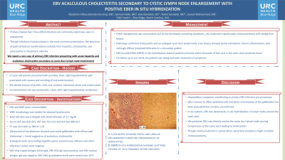
Initial management was conservative but as she developed worsening symptoms, she underwent laparoscopic cholecystectomy with wedge liver biopsy. Pathology confirmed cholecystitis and an enlarged cystic duct lymph node. Liver biopsy showed portal and lobular chronic inflammation, with strikingly diffuse lymphoid infiltration in a sinusoidal pattern (Fig 1). EBV-encoded RNA (EBER) in situ hybridization stained positive primarily within sinusoids of liver and in the cystic duct lymphoid tissue. On follow up at one week, the patient was doing well with resolution of symptoms.
Discussion: Hepatobiliary symptoms manifesting as primary EBV infection are uncommon. EBV is known to affect epithelial cells and direct viral invasion of the gallbladder has been postulated but remains unconfirmed. In our patient, EBV was detected by in situ hybridization in lymph nodes around the cystic duct. We postulate EBV may directly involve the cystic duct lymph node causing compression of the cystic duct leading to cholecystitis. Though mostly treatment is conservative, persistent symptoms might mandate cholecystectomy.

Disclosures:
Madeline Vithya Barnaba Durairaj, MBBS, MD, DM, Samuel Addo, MD, Kyle Burnette, DO, Rahul Sampath, MD, Suneel Mohammed, MD. P2953 - EBV Acalculous Cholecystitis Secondary to Cystic Lymph Node Enlargement With Positive EBER in situ Hybridization, ACG 2023 Annual Scientific Meeting Abstracts. Vancouver, BC, Canada: American College of Gastroenterology.
UNC Health Blue Ridge, Morganton, NC
Introduction: Primary Epstein-Barr Virus (EBV) infections are commonly subclinical, seen in adolescents. Though infectious mononucleosis is the most common presentation, the spectrum of gastrointestinal manifestations extends from hepatitis, cholecystitis, and pancreatitis to mesenteric adenitis. We report a rare case of primary EBV infection presenting with acute hepatitis and acalculous cholecystitis secondary to cystic duct lymph node involvement.
Case Description/Methods: A 27-year-old woman presented with jaundice, fever, right hypochondriac pain associated with nausea and vomiting of one week duration. She denied history of pruritis, rash, sick contacts, substance abuse and recent travel. On examination she was tachycardic, icteric with right hypochondriac tenderness. CBC and BMP were unremarkable. WBC morphology was notable for atypical lymphocytes. Total bilirubin was 5.4 mg/dl with direct bilirubin of 3.7 mg/dl. Serum ALT was 810 IU/L, AST was 751 IU/L and ALP was 486 IU/L. PT was 14.4 with an INR of 1.10. Ultrasound of the abdomen showed contracted gallbladder with diffuse wall thickening ( > 5mm) suggestive of acalculous cholecystitis. Etiological work up including hepatitis panel, autoimmune, Wilsons and other infectious causes were negative. EBV Viral Capsid Antigen (VCA) IgM, EBV VCA IgG were positive, and EBV nuclear antigen IgG was negative. EBV DNA quantitative levels were noted to be 3377.
Initial management was conservative but as she developed worsening symptoms, she underwent laparoscopic cholecystectomy with wedge liver biopsy. Pathology confirmed cholecystitis and an enlarged cystic duct lymph node. Liver biopsy showed portal and lobular chronic inflammation, with strikingly diffuse lymphoid infiltration in a sinusoidal pattern (Fig 1). EBV-encoded RNA (EBER) in situ hybridization stained positive primarily within sinusoids of liver and in the cystic duct lymphoid tissue. On follow up at one week, the patient was doing well with resolution of symptoms.
Discussion: Hepatobiliary symptoms manifesting as primary EBV infection are uncommon. EBV is known to affect epithelial cells and direct viral invasion of the gallbladder has been postulated but remains unconfirmed. In our patient, EBV was detected by in situ hybridization in lymph nodes around the cystic duct. We postulate EBV may directly involve the cystic duct lymph node causing compression of the cystic duct leading to cholecystitis. Though mostly treatment is conservative, persistent symptoms might mandate cholecystectomy.

Figure: A: Liver biopsy showing portal and lobular inflammation consisting predominantly of lymphocytes.
B: EBER In situ hybridization showing scattered staining of cells primarily within sinusoids.
B: EBER In situ hybridization showing scattered staining of cells primarily within sinusoids.
Disclosures:
Madeline Vithya Barnaba Durairaj indicated no relevant financial relationships.
Samuel Addo indicated no relevant financial relationships.
Kyle Burnette indicated no relevant financial relationships.
Rahul Sampath indicated no relevant financial relationships.
Suneel Mohammed indicated no relevant financial relationships.
Madeline Vithya Barnaba Durairaj, MBBS, MD, DM, Samuel Addo, MD, Kyle Burnette, DO, Rahul Sampath, MD, Suneel Mohammed, MD. P2953 - EBV Acalculous Cholecystitis Secondary to Cystic Lymph Node Enlargement With Positive EBER in situ Hybridization, ACG 2023 Annual Scientific Meeting Abstracts. Vancouver, BC, Canada: American College of Gastroenterology.
