Tuesday Poster Session
Category: Liver
P3957 - Diabetic Hepatosclerosis: A Rare Complication of a Common Disease
Tuesday, October 24, 2023
10:30 AM - 4:00 PM PT
Location: Exhibit Hall

Has Audio

Baha Aldeen Bani Fawwaz, MD
AdventHealth
Orlando, FL
Presenting Author(s)
Baha Fawwaz, MD, Vishwas Vanar, MD
AdventHealth, Orlando, FL
Introduction: Diabetes mellitus is a set of metabolic disorders that lead to elevated blood sugar levels as a result of inadequate insulin production or insulin resistance. These disorders can cause complications that affect nearly every system in the body. Among the most common gastrointestinal complications of DM are non-alcoholic fatty liver disease (NAFLD), gastroparesis, and gastroesophageal reflux disease. Diabetic hepatosclerosis (DHS) is a recently recognized form of diabetic microangiopathy, which is characterized by presinusoidal fibrosis. We present a case of DHS in a patient with long-standing type 1 DM, which was confirmed by liver biopsy.
Case Description/Methods: A 39-year-old female patient with poorly controlled type 1 DM,ESRD on hemodialysis,hypertension,and recurrent skin abscesses presented with scleral icterus.She had been experiencing chronic watery brown diarrhea, nausea, and generalized weakness for the past month, as well as multiple furuncles on her body and scalp. Examination revealed scleral icterus and a large fluctuant swelling (12cm x 12cm) on her scalp. Labs are shown in table 1.
Despite a comprehensive liver disease workup,no significant abnormalities were found, and a liver biopsy was performed, which revealed extensive perisinusoidal and portal fibrosis that may be indicative of DHS.The patient was started on ursodiol,and her total bilirubin and elevated alkaline phosphatase (ALP) levels began to trend downwards, while her diarrhea improved.
During her hospital stay,the patient underwent surgical drainage for her abscess.
Discussion: The most common hepatic pathology associated with DM is NAFLD,which is characterized by the accumulation of lipids in the liver. Another form of liver damage associated with DM is fibrosis, which is thought to be a type of microangiopathy.In diabetic microangiopathy, capillary basement membrane (BM) thickening is the histological hallmark.In DHS, perisinusoidal fibrosis forms BM around the sinusoids since they do not have their BM.This process is known as the capillarization of sinusoids. Unlike NAFLD, which is associated with elevated AST/ALT levels, DHS is often characterized by ALP levels.DHS is more common in patients with long-standing type 1 DM and end-organ complications.Although there are no established guidelines for managing DHS, ursodiol was used to successfully improve the patient's symptoms in this case.Despite DHS being a non-cirrhotic disease, further long-term follow-up is necessary to better understand its clinical significance.

Disclosures:
Baha Fawwaz, MD, Vishwas Vanar, MD. P3957 - Diabetic Hepatosclerosis: A Rare Complication of a Common Disease, ACG 2023 Annual Scientific Meeting Abstracts. Vancouver, BC, Canada: American College of Gastroenterology.
AdventHealth, Orlando, FL
Introduction: Diabetes mellitus is a set of metabolic disorders that lead to elevated blood sugar levels as a result of inadequate insulin production or insulin resistance. These disorders can cause complications that affect nearly every system in the body. Among the most common gastrointestinal complications of DM are non-alcoholic fatty liver disease (NAFLD), gastroparesis, and gastroesophageal reflux disease. Diabetic hepatosclerosis (DHS) is a recently recognized form of diabetic microangiopathy, which is characterized by presinusoidal fibrosis. We present a case of DHS in a patient with long-standing type 1 DM, which was confirmed by liver biopsy.
Case Description/Methods: A 39-year-old female patient with poorly controlled type 1 DM,ESRD on hemodialysis,hypertension,and recurrent skin abscesses presented with scleral icterus.She had been experiencing chronic watery brown diarrhea, nausea, and generalized weakness for the past month, as well as multiple furuncles on her body and scalp. Examination revealed scleral icterus and a large fluctuant swelling (12cm x 12cm) on her scalp. Labs are shown in table 1.
Despite a comprehensive liver disease workup,no significant abnormalities were found, and a liver biopsy was performed, which revealed extensive perisinusoidal and portal fibrosis that may be indicative of DHS.The patient was started on ursodiol,and her total bilirubin and elevated alkaline phosphatase (ALP) levels began to trend downwards, while her diarrhea improved.
During her hospital stay,the patient underwent surgical drainage for her abscess.
Discussion: The most common hepatic pathology associated with DM is NAFLD,which is characterized by the accumulation of lipids in the liver. Another form of liver damage associated with DM is fibrosis, which is thought to be a type of microangiopathy.In diabetic microangiopathy, capillary basement membrane (BM) thickening is the histological hallmark.In DHS, perisinusoidal fibrosis forms BM around the sinusoids since they do not have their BM.This process is known as the capillarization of sinusoids. Unlike NAFLD, which is associated with elevated AST/ALT levels, DHS is often characterized by ALP levels.DHS is more common in patients with long-standing type 1 DM and end-organ complications.Although there are no established guidelines for managing DHS, ursodiol was used to successfully improve the patient's symptoms in this case.Despite DHS being a non-cirrhotic disease, further long-term follow-up is necessary to better understand its clinical significance.

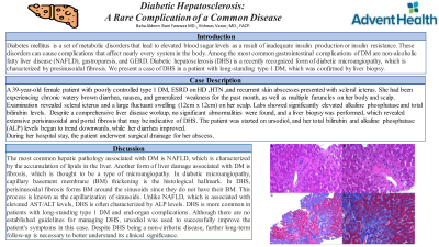
Figure: Image (A): representative portal tract with a bile duct, arteriole and portal venule. You can appreciate a subtle pink staining material around some of the hepatocytes which represents collagen. There is also some brown granular material which is iron in Kupffer cells.
Image (B): representative image of centrilobular area showing a terminal hepatic venule. There is significant collagen accumulation around this vessel and also around some of the hepatocytes in the area (pink homogenous material)
Image (C): Trichrome stain, low power, showing an intricate pattern of collagen around individual hepatocytes. The collagen stains blue and the hepatocytes are red.
Image (D): Trichrome stain, high power showing the intricate “chicken wire” pattern of collagen around individual cells.
Disclosures:
Baha Fawwaz indicated no relevant financial relationships.
Vishwas Vanar indicated no relevant financial relationships.
Baha Fawwaz, MD, Vishwas Vanar, MD. P3957 - Diabetic Hepatosclerosis: A Rare Complication of a Common Disease, ACG 2023 Annual Scientific Meeting Abstracts. Vancouver, BC, Canada: American College of Gastroenterology.
