Tuesday Poster Session
Category: Biliary/Pancreas
P2961 - Chasing Comets: A Case of Recurrent Intrahepatic Stones
Tuesday, October 24, 2023
10:30 AM - 4:00 PM PT
Location: Exhibit Hall

Has Audio
- PK
Pradeep Koripella, MD
Kaiser Permanente
San Francisco, CA
Presenting Author(s)
Pradeep Koripella, MD1, Lawrence Leung, MD, MPH1, Nizar A. Mukhtar, MD2, Christopher Hamerski, MD1
1Kaiser Permanente, San Francisco, CA; 2Kaiser Permanente San Francisco Medical Center, San Francisco, CA
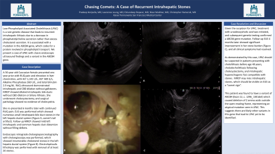
Introduction: Low-Phospholipid Associated Cholelithiasis (LPAC) is a rare genetic disease that leads to recurrent intrahepatic lithiasis due to a decrease in phosphatidylcholine secretion rather than excess cholesterol secretion. It is associated with a mutation in the ABCB4 gene, which codes for a protein involved in phospholipid transport. We present a case of LPAC with classic endoscopic ultrasound findings and a variant in the ABCB4 gene.
Case Description/Methods: A 30-year-old Caucasian female presented one year prior with right upper quadrant (RUQ) pain and elevation in liver chemistries, with ALT 1,142 U/L, AST 589 U/L, Alkaline Phosphatase 260 U/L, and total bilirubin 2.9 mg/dL. RUQ ultrasound demonstrated intrahepatic and common bile duct (CBD) dilation without gallstones. Magnetic resonance cholangiopancreatography (MRCP) showed dilated intrahepatic bile ducts without CBD dilation or biliary lithiasis. She underwent cholecystectomy, and surgical pathology showed no evidence of cholecystitis.
She re-presented 6 months later with continued RUQ pain. Endoscopic ultrasound was performed which showed numerous small (2-7mm) intrahepatic bile duct stones in the left hepatic ductal system (Figure A, comet’s tail artifact). Follow up MRCP showed mild left intrahepatic and common hepatic duct distention without filling defects. Endoscopic retrograde cholangiopancreatography with cholangioscopy was performed, which showed innumerable cholesterol stones in the left hepatic ductal system (Figure B). Electrohydraulic lithotripsy was performed with removal of at least 20 stones.
Given the suspicion for LPAC, treatment with ursodeoxycholic acid (UDCA) was initiated, and subsequent genetic testing confirmed a ABCB4 gene mutation. Follow-up EUS 4 months later showed significant improvement in her stone burden (Figure C), and all clinical symptoms had resolved.
Discussion: As demonstrated by this case, LPAC should be suspected in patients presenting with cholelithiasis before age 40 years, choledocholithiasis following cholecystectomy, and intrahepatic hyperechogenic foci compatible with stones. MRCP may miss intrahepatic stones, which should be visible on EUS as a “comet sign”.
This patient was found to have a variant of ABCB4 (Exon 12, c. 1296_1301del) which caused deletion of 2 amino acids outside the open reading frame, representing an atypical mutation seen in LPAC. This suggests there are likely other variants in this gene that lead to LPAC yet to be identified.

Disclosures:
Pradeep Koripella, MD1, Lawrence Leung, MD, MPH1, Nizar A. Mukhtar, MD2, Christopher Hamerski, MD1. P2961 - Chasing Comets: A Case of Recurrent Intrahepatic Stones, ACG 2023 Annual Scientific Meeting Abstracts. Vancouver, BC, Canada: American College of Gastroenterology.
1Kaiser Permanente, San Francisco, CA; 2Kaiser Permanente San Francisco Medical Center, San Francisco, CA
Introduction: Low-Phospholipid Associated Cholelithiasis (LPAC) is a rare genetic disease that leads to recurrent intrahepatic lithiasis due to a decrease in phosphatidylcholine secretion rather than excess cholesterol secretion. It is associated with a mutation in the ABCB4 gene, which codes for a protein involved in phospholipid transport. We present a case of LPAC with classic endoscopic ultrasound findings and a variant in the ABCB4 gene.
Case Description/Methods: A 30-year-old Caucasian female presented one year prior with right upper quadrant (RUQ) pain and elevation in liver chemistries, with ALT 1,142 U/L, AST 589 U/L, Alkaline Phosphatase 260 U/L, and total bilirubin 2.9 mg/dL. RUQ ultrasound demonstrated intrahepatic and common bile duct (CBD) dilation without gallstones. Magnetic resonance cholangiopancreatography (MRCP) showed dilated intrahepatic bile ducts without CBD dilation or biliary lithiasis. She underwent cholecystectomy, and surgical pathology showed no evidence of cholecystitis.
She re-presented 6 months later with continued RUQ pain. Endoscopic ultrasound was performed which showed numerous small (2-7mm) intrahepatic bile duct stones in the left hepatic ductal system (Figure A, comet’s tail artifact). Follow up MRCP showed mild left intrahepatic and common hepatic duct distention without filling defects. Endoscopic retrograde cholangiopancreatography with cholangioscopy was performed, which showed innumerable cholesterol stones in the left hepatic ductal system (Figure B). Electrohydraulic lithotripsy was performed with removal of at least 20 stones.
Given the suspicion for LPAC, treatment with ursodeoxycholic acid (UDCA) was initiated, and subsequent genetic testing confirmed a ABCB4 gene mutation. Follow-up EUS 4 months later showed significant improvement in her stone burden (Figure C), and all clinical symptoms had resolved.
Discussion: As demonstrated by this case, LPAC should be suspected in patients presenting with cholelithiasis before age 40 years, choledocholithiasis following cholecystectomy, and intrahepatic hyperechogenic foci compatible with stones. MRCP may miss intrahepatic stones, which should be visible on EUS as a “comet sign”.
This patient was found to have a variant of ABCB4 (Exon 12, c. 1296_1301del) which caused deletion of 2 amino acids outside the open reading frame, representing an atypical mutation seen in LPAC. This suggests there are likely other variants in this gene that lead to LPAC yet to be identified.

Figure: A) Endoscopic ultrasound showing a "comet's tail" artifact, suggesting biliary lithiasis
B) Cholangioscopy demonstrating cholesterol stones
C) Endoscopic ultrasound demonstrating resolution of previously seen artifacts after treatment (signifies improvement in stone burden)
B) Cholangioscopy demonstrating cholesterol stones
C) Endoscopic ultrasound demonstrating resolution of previously seen artifacts after treatment (signifies improvement in stone burden)
Disclosures:
Pradeep Koripella indicated no relevant financial relationships.
Lawrence Leung indicated no relevant financial relationships.
Nizar Mukhtar indicated no relevant financial relationships.
Christopher Hamerski indicated no relevant financial relationships.
Pradeep Koripella, MD1, Lawrence Leung, MD, MPH1, Nizar A. Mukhtar, MD2, Christopher Hamerski, MD1. P2961 - Chasing Comets: A Case of Recurrent Intrahepatic Stones, ACG 2023 Annual Scientific Meeting Abstracts. Vancouver, BC, Canada: American College of Gastroenterology.
