Tuesday Poster Session
Category: Biliary/Pancreas
P2965 - An Unusual Case of Renal Cell Carcinoma Metastasizing to the Pancreas
Tuesday, October 24, 2023
10:30 AM - 4:00 PM PT
Location: Exhibit Hall

Has Audio

Robert Schoeneich
Advocate Lutheran General Hospital
Coralville, IA
Presenting Author(s)
Robert Schoeneich, 1, David H. Kruchko, DO2, Kenneth D.. Chi, MD3
1Advocate Lutheran General Hospital, Wrightstown, WI; 2Advocate Aurora Health, Chicago, IL; 3Advocate Lutheran General Hospital, Glenview, IL
Introduction: Renal cell carcinoma (RCC) has an estimated incidence of 81,800 for 2023 in the United States, affecting men to women in a 2:1 ratio [1]. RCC metastasizes in about 20-30% of cases, with the most common sites being the lungs, bones, liver and brain [3]. Metastasis to the pancreas is very rare, accounting for only 2-5% of all malignancies affecting the pancreas [2]. We present a rare case of RCC metastasis to the pancreas in a female patient 6 years after initial diagnosis and treatment of RCC.
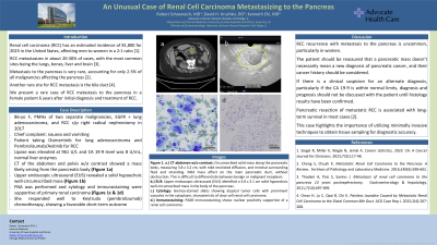
Case Description/Methods: An 86-year-old female with a past medical history of two separate malignancies in her lifetime, a remote diagnosis of EGFR (epidermal growth factor receptor) positive lung adenocarcinoma, and more recently diagnosed with RCC s/p right radical nephrectomy in 2017, presented with nausea and vomiting. At the time of presentation, she was taking Osimertinib for lung adenocarcinoma and Pembrolizumab/Axitinib for RCC. Liver enzymes were within normal limits, and she had not noticed any jaundice prior to admission. Her lipase was elevated at 961 U/L and her CA 19-9 level was 8 U/mL. CT of the abdomen and pelvis without contrast revealed a mass likely arising from the pancreatic body, prompting GI consult. Minimal surrounding fluid and stranding was noted with mild pancreatic duct dilation but no evidence of pancreatitis (Figure 1a). Upper endoscopic ultrasound (EUS) revealed a solid hypoechoic well-circumscribed mass with additional findings described below (Figure 1b). FNA was performed and cytology and immunostaining were supportive of primary renal carcinoma (Figure 1c & 1d). The patient responded well to Keytruda chemotherapy, showing a favorable short-term outcome.
Discussion: RCC recurrence with metastasis to the pancreas is uncommon, particularly in women. The patient should be reassured that a pancreatic mass doesn’t necessarily mean a new diagnosis of pancreatic cancer. If there is a clinical suspicion for an alternate diagnosis, particularly if the CA 19-9 is within normal limits, diagnosis and prognosis should not be discussed with the patient until histology results have been confirmed.
References
1. Siegel R, Miller K, Wagle N, Jemal A. CA: A Cancer Journal for Clinicians. 2023;73(1):17-48.
2. Cheng S, Chuah K. Archives of Pathology and Laboratory Medicine. 2016;140(6):598-602.
3. Thadani A, Pais S, Savino J. Gastroenterology & hepatology. 2011;7(10):697-699.

Disclosures:
Robert Schoeneich, 1, David H. Kruchko, DO2, Kenneth D.. Chi, MD3. P2965 - An Unusual Case of Renal Cell Carcinoma Metastasizing to the Pancreas, ACG 2023 Annual Scientific Meeting Abstracts. Vancouver, BC, Canada: American College of Gastroenterology.
1Advocate Lutheran General Hospital, Wrightstown, WI; 2Advocate Aurora Health, Chicago, IL; 3Advocate Lutheran General Hospital, Glenview, IL
Introduction: Renal cell carcinoma (RCC) has an estimated incidence of 81,800 for 2023 in the United States, affecting men to women in a 2:1 ratio [1]. RCC metastasizes in about 20-30% of cases, with the most common sites being the lungs, bones, liver and brain [3]. Metastasis to the pancreas is very rare, accounting for only 2-5% of all malignancies affecting the pancreas [2]. We present a rare case of RCC metastasis to the pancreas in a female patient 6 years after initial diagnosis and treatment of RCC.
Case Description/Methods: An 86-year-old female with a past medical history of two separate malignancies in her lifetime, a remote diagnosis of EGFR (epidermal growth factor receptor) positive lung adenocarcinoma, and more recently diagnosed with RCC s/p right radical nephrectomy in 2017, presented with nausea and vomiting. At the time of presentation, she was taking Osimertinib for lung adenocarcinoma and Pembrolizumab/Axitinib for RCC. Liver enzymes were within normal limits, and she had not noticed any jaundice prior to admission. Her lipase was elevated at 961 U/L and her CA 19-9 level was 8 U/mL. CT of the abdomen and pelvis without contrast revealed a mass likely arising from the pancreatic body, prompting GI consult. Minimal surrounding fluid and stranding was noted with mild pancreatic duct dilation but no evidence of pancreatitis (Figure 1a). Upper endoscopic ultrasound (EUS) revealed a solid hypoechoic well-circumscribed mass with additional findings described below (Figure 1b). FNA was performed and cytology and immunostaining were supportive of primary renal carcinoma (Figure 1c & 1d). The patient responded well to Keytruda chemotherapy, showing a favorable short-term outcome.
Discussion: RCC recurrence with metastasis to the pancreas is uncommon, particularly in women. The patient should be reassured that a pancreatic mass doesn’t necessarily mean a new diagnosis of pancreatic cancer. If there is a clinical suspicion for an alternate diagnosis, particularly if the CA 19-9 is within normal limits, diagnosis and prognosis should not be discussed with the patient until histology results have been confirmed.
References
1. Siegel R, Miller K, Wagle N, Jemal A. CA: A Cancer Journal for Clinicians. 2023;73(1):17-48.
2. Cheng S, Chuah K. Archives of Pathology and Laboratory Medicine. 2016;140(6):598-602.
3. Thadani A, Pais S, Savino J. Gastroenterology & hepatology. 2011;7(10):697-699.

Figure: Figure 1. a.) CT abdomen w/o contrast: Circumscribed solid mass along the pancreatic body, measuring 3.8 x 3.2 cm, with mild restricted diffusion, and minimal surrounding fluid and stranding. Mild mass effect on the main pancreatic duct, without obstruction. This is difficult to differentiate between benign or malignant neoplasm. b.) EUS: Upper endoscopic ultrasound (EUS) identified a 3.8 x 3.1 cm solid hypoechoic well-circumscribed mass in the body of the pancreas. c.) Cytology: Giemsa-stained slides showing atypical tumor cells with prominent vacuoles in the cytoplasm, characteristic of clear cell renal cell carcinoma. d.) Immunostaining: PAX8 immunostaining shows nuclear positivity supportive of a renal cell carcinoma.
Disclosures:
Robert Schoeneich indicated no relevant financial relationships.
David Kruchko indicated no relevant financial relationships.
Kenneth Chi: Boston Scientific – Consultant.
Robert Schoeneich, 1, David H. Kruchko, DO2, Kenneth D.. Chi, MD3. P2965 - An Unusual Case of Renal Cell Carcinoma Metastasizing to the Pancreas, ACG 2023 Annual Scientific Meeting Abstracts. Vancouver, BC, Canada: American College of Gastroenterology.
