Tuesday Poster Session
Category: Biliary/Pancreas
P2966 - Unusual and Elusive: Investigating an Uncommon Hemosuccus Pancreaticus Case
Tuesday, October 24, 2023
10:30 AM - 4:00 PM PT
Location: Exhibit Hall

Has Audio
- BS
Balarama K. Surapaneni, MBBS
University of Iowa Hospitals & Clinics
Iowa City, IA
Presenting Author(s)
Balarama K. Surapaneni, MBBS, Aniket Mittal, MBBS, Taraz Imani. Ramos, MD, Samragnyi Madala, MBBS, Rintaro Hashimoto, MD, PhD
University of Iowa Hospitals & Clinics, Iowa City, IA
Introduction: Splenic artery is the third most common cause of intrabdominal aneurysms and most common aneurysms and most common visceral aneurysms . We present a rare case of splenic artery pseudoaneurysm in association with hereditary pancreatitis and with bleeding in the pancreatic duct leading to Hemosuccus pancreaticus( HP) in a patient with associated segmental PE.
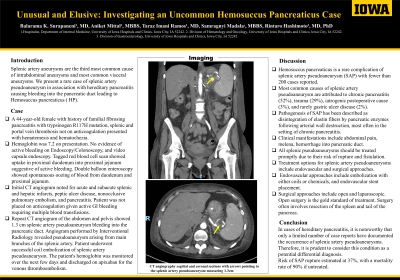
Case Description/Methods: A 44-year-old female with history of familial fibrosing pancreatitis with trypsinogen R117H mutation, splenic and portal vein thrombosis no longer on anticoagulation, chronic depression, anxiety, chronic pain, and chronic hidradenitis who presented with chief complaints of ongoing hematemesis and hematochezia. Laboratory work during admission noted for Hemoglobin 7.2, hematocrit 25. Gastroenterology (GI) was consulted and underwent Endoscopy/Colonoscopy which was negative for active or recent bleeding sources. Patient later underwent video capsule endoscopy which was also inconclusive for acute bleed. The patient had ongoing bloody bowel movements and required frequent blood transfusions. Tagged RBC scan showed uptake in proximal duodenum into proximal jejunum suggestive of active bleeding. Later underwent double balloon study showed spontaneous oozing of blood from duodenum and proximal jejunum. Initial CTA noted for acute and subacute splenic infarcts, hepatic infarct, peptic ulcer disease, pulmonary embolism, and pancreatitis. CTA chest was noted for Nonocclusive thromboembolic pulmonary disease involving the right lower lobe segmental artery. Bilateral lower extremity duplex showed no evidence of DVT. Patient was not placed on anticoagulation given recurrent GI bleeding with dropping hemoglobin and frequent blood transfusions. The second CTA abdomen and pelvis ordered was noted for showing 1.3 cm (about 0.51 in) splenic artery pseudoaneurysm into the pancreatic duct with acute and subacute splenic infarcts, hepatic infarct, peptic ulcer disease and pancreatitis. Interventional Radiology was consulted, and an angiogram was planned for splenic artery pseudoaneurysm. Angiogram revealed pseudoaneurysm arising from one of the main branches of the splenic artery. Patient underwent Successful coil embolization of splenic artery pseudoaneurysm which was completely excluded on post treatment angiogram. The patient's hemoglobin was monitored for a couple of days and remained stable and switched to apixaban and was discharged home.
Discussion: Hemosuccus pancreaticus is a rare complication of Splenic artery pseudoaneurysm.

Disclosures:
Balarama K. Surapaneni, MBBS, Aniket Mittal, MBBS, Taraz Imani. Ramos, MD, Samragnyi Madala, MBBS, Rintaro Hashimoto, MD, PhD. P2966 - Unusual and Elusive: Investigating an Uncommon Hemosuccus Pancreaticus Case, ACG 2023 Annual Scientific Meeting Abstracts. Vancouver, BC, Canada: American College of Gastroenterology.
University of Iowa Hospitals & Clinics, Iowa City, IA
Introduction: Splenic artery is the third most common cause of intrabdominal aneurysms and most common aneurysms and most common visceral aneurysms . We present a rare case of splenic artery pseudoaneurysm in association with hereditary pancreatitis and with bleeding in the pancreatic duct leading to Hemosuccus pancreaticus( HP) in a patient with associated segmental PE.
Case Description/Methods: A 44-year-old female with history of familial fibrosing pancreatitis with trypsinogen R117H mutation, splenic and portal vein thrombosis no longer on anticoagulation, chronic depression, anxiety, chronic pain, and chronic hidradenitis who presented with chief complaints of ongoing hematemesis and hematochezia. Laboratory work during admission noted for Hemoglobin 7.2, hematocrit 25. Gastroenterology (GI) was consulted and underwent Endoscopy/Colonoscopy which was negative for active or recent bleeding sources. Patient later underwent video capsule endoscopy which was also inconclusive for acute bleed. The patient had ongoing bloody bowel movements and required frequent blood transfusions. Tagged RBC scan showed uptake in proximal duodenum into proximal jejunum suggestive of active bleeding. Later underwent double balloon study showed spontaneous oozing of blood from duodenum and proximal jejunum. Initial CTA noted for acute and subacute splenic infarcts, hepatic infarct, peptic ulcer disease, pulmonary embolism, and pancreatitis. CTA chest was noted for Nonocclusive thromboembolic pulmonary disease involving the right lower lobe segmental artery. Bilateral lower extremity duplex showed no evidence of DVT. Patient was not placed on anticoagulation given recurrent GI bleeding with dropping hemoglobin and frequent blood transfusions. The second CTA abdomen and pelvis ordered was noted for showing 1.3 cm (about 0.51 in) splenic artery pseudoaneurysm into the pancreatic duct with acute and subacute splenic infarcts, hepatic infarct, peptic ulcer disease and pancreatitis. Interventional Radiology was consulted, and an angiogram was planned for splenic artery pseudoaneurysm. Angiogram revealed pseudoaneurysm arising from one of the main branches of the splenic artery. Patient underwent Successful coil embolization of splenic artery pseudoaneurysm which was completely excluded on post treatment angiogram. The patient's hemoglobin was monitored for a couple of days and remained stable and switched to apixaban and was discharged home.
Discussion: Hemosuccus pancreaticus is a rare complication of Splenic artery pseudoaneurysm.

Figure: 1.3 cm (about 0.51 in) splenic artery pseudoaneurysm into the pancreatic duct
Disclosures:
Balarama Surapaneni indicated no relevant financial relationships.
Aniket Mittal indicated no relevant financial relationships.
Taraz Ramos indicated no relevant financial relationships.
Samragnyi Madala indicated no relevant financial relationships.
Rintaro Hashimoto indicated no relevant financial relationships.
Balarama K. Surapaneni, MBBS, Aniket Mittal, MBBS, Taraz Imani. Ramos, MD, Samragnyi Madala, MBBS, Rintaro Hashimoto, MD, PhD. P2966 - Unusual and Elusive: Investigating an Uncommon Hemosuccus Pancreaticus Case, ACG 2023 Annual Scientific Meeting Abstracts. Vancouver, BC, Canada: American College of Gastroenterology.
