Tuesday Poster Session
Category: Biliary/Pancreas
P2969 - Secondary Sclerosing Cholangitis After Antibiotic-Related Drug-Induced Liver Injury
Tuesday, October 24, 2023
10:30 AM - 4:00 PM PT
Location: Exhibit Hall

Has Audio

Emilie S. Kim, MD
St. Luke’s University Hospital
Bethlehem, PA
Presenting Author(s)
Emilie S. Kim, MD1, Janak Bahirwani, MD2, Brian Kim, DO1, Liyan Xu, MD1, Matthew A. Glavin, MD2, Vishal Patel, MD1
1St. Luke’s University Hospital, Bethlehem, PA; 2St. Luke's University Hospital, Bethlehem, PA
Introduction: Literature on secondary sclerosing cholangitis (SSC) due to drug-induced liver injury (DILI) is limited to case studies of antineoplastic immunotherapy, herbal drugs, sevofluorane, and moxifloxacin. We present a case of SSC after DILI from amoxicillin-clavulanate, metronidazole, and ceftriaxone use.
Case Description/Methods: A 58-year-old male admitted for diabetic ketoacidosis and acalculous cholecystitis received percutaneous cholecystostomy and fluid culture grew klebsiella, enterococcus, and E. Coli. He was treated with amoxicillin-clavulanate, metronidazole and ceftriaxone. Liver function tests(LFT) showed total bilirubin (2.84 mg/dl), ALP (285 IU/L), AST (192 IU/L), and ALT (140 IU/L) which peaked and stabilized. After one month, he presented with jaundice and elevated LFT with rising bilirubin and LFT’s. RUCAM score 6 pointed to amoxicillin-clavulanate as the probable cause for liver injury. Magnetic resonance cholangiopancreatography (MRCP) and Endoscopic retrograde cholangiopancreatography (ERCP) did not show any stone, dilation, stricture or mass. His first liver biopsy showed active hepatitis, cholestasis, and no fibrosis, consistent with DILI from antibiotic exposure.
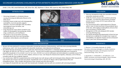
Four months later, MRCP revealed obstruction of left hepatic duct with abrupt cutoff and intrahepatic biliary dilatation (Fig. 1a, 1c). ERCP showed left-main hepatic duct stricture and stent was placed. Total bilirubin rose to a peak of 19. Biopsy ruled out IgG4 disease (PSC) and cholangiocarcinoma. Chronic liver disease etiology testing were negative. He received a 4-month taper of steroids with modest improvement in bilirubin. 11 months from initial injury, liver biopsy showed cholestasis, bile duct injury with focal lymphocytic cholangitis, and duct loss with scar, suggestive for sclerosing cholangitis(Fig. 1b, 1c). He developed decompensated cirrhosis but deemed not a candidate for transplant due to age and comorbidities. He opted for hospice and passed away 2 years from initial presentation.
Discussion: Only 3 cases from one retrospective study cites association between amoxicillin-clavulanate and SSC. This case is unique due to the rapid progression of sclerosing cholangitis with imaging revealing biliary strictures that were not present 5 months earlier. Management of DILI and subsequent SSC includes removing offending drug. This highlights the rapid progression of SSC toward end-stage liver cirrhosis within 1 year of initial insult delineating the importance of early identification SSC due to antibiotics.

Disclosures:
Emilie S. Kim, MD1, Janak Bahirwani, MD2, Brian Kim, DO1, Liyan Xu, MD1, Matthew A. Glavin, MD2, Vishal Patel, MD1. P2969 - Secondary Sclerosing Cholangitis After Antibiotic-Related Drug-Induced Liver Injury, ACG 2023 Annual Scientific Meeting Abstracts. Vancouver, BC, Canada: American College of Gastroenterology.
1St. Luke’s University Hospital, Bethlehem, PA; 2St. Luke's University Hospital, Bethlehem, PA
Introduction: Literature on secondary sclerosing cholangitis (SSC) due to drug-induced liver injury (DILI) is limited to case studies of antineoplastic immunotherapy, herbal drugs, sevofluorane, and moxifloxacin. We present a case of SSC after DILI from amoxicillin-clavulanate, metronidazole, and ceftriaxone use.
Case Description/Methods: A 58-year-old male admitted for diabetic ketoacidosis and acalculous cholecystitis received percutaneous cholecystostomy and fluid culture grew klebsiella, enterococcus, and E. Coli. He was treated with amoxicillin-clavulanate, metronidazole and ceftriaxone. Liver function tests(LFT) showed total bilirubin (2.84 mg/dl), ALP (285 IU/L), AST (192 IU/L), and ALT (140 IU/L) which peaked and stabilized. After one month, he presented with jaundice and elevated LFT with rising bilirubin and LFT’s. RUCAM score 6 pointed to amoxicillin-clavulanate as the probable cause for liver injury. Magnetic resonance cholangiopancreatography (MRCP) and Endoscopic retrograde cholangiopancreatography (ERCP) did not show any stone, dilation, stricture or mass. His first liver biopsy showed active hepatitis, cholestasis, and no fibrosis, consistent with DILI from antibiotic exposure.
Four months later, MRCP revealed obstruction of left hepatic duct with abrupt cutoff and intrahepatic biliary dilatation (Fig. 1a, 1c). ERCP showed left-main hepatic duct stricture and stent was placed. Total bilirubin rose to a peak of 19. Biopsy ruled out IgG4 disease (PSC) and cholangiocarcinoma. Chronic liver disease etiology testing were negative. He received a 4-month taper of steroids with modest improvement in bilirubin. 11 months from initial injury, liver biopsy showed cholestasis, bile duct injury with focal lymphocytic cholangitis, and duct loss with scar, suggestive for sclerosing cholangitis(Fig. 1b, 1c). He developed decompensated cirrhosis but deemed not a candidate for transplant due to age and comorbidities. He opted for hospice and passed away 2 years from initial presentation.
Discussion: Only 3 cases from one retrospective study cites association between amoxicillin-clavulanate and SSC. This case is unique due to the rapid progression of sclerosing cholangitis with imaging revealing biliary strictures that were not present 5 months earlier. Management of DILI and subsequent SSC includes removing offending drug. This highlights the rapid progression of SSC toward end-stage liver cirrhosis within 1 year of initial insult delineating the importance of early identification SSC due to antibiotics.

Figure: Figure 1. (a) segmental stricutre of left hepatic duct with wall thickening on MRCP, (b) bile duct injury including focal lymphocytic cholangitis on second liver biopsy 11 months after initial presentation, (c) segmental stricture of left hepatic duct with diffuse upstream left intra-hepatic duct dilatation enhancement on MRCP 5 months after initial presentation, and (d) duct loss with scar and bile ductular reaction on liver biopsy 11 months after initial presentation
Disclosures:
Emilie S. Kim indicated no relevant financial relationships.
Janak Bahirwani indicated no relevant financial relationships.
Brian Kim indicated no relevant financial relationships.
Liyan Xu indicated no relevant financial relationships.
Matthew A. Glavin indicated no relevant financial relationships.
Vishal Patel indicated no relevant financial relationships.
Emilie S. Kim, MD1, Janak Bahirwani, MD2, Brian Kim, DO1, Liyan Xu, MD1, Matthew A. Glavin, MD2, Vishal Patel, MD1. P2969 - Secondary Sclerosing Cholangitis After Antibiotic-Related Drug-Induced Liver Injury, ACG 2023 Annual Scientific Meeting Abstracts. Vancouver, BC, Canada: American College of Gastroenterology.
