Tuesday Poster Session
Category: Biliary/Pancreas
P2973 - Rare Case of a Pancreatic Sclerosing Hemangioma
Tuesday, October 24, 2023
10:30 AM - 4:00 PM PT
Location: Exhibit Hall

Has Audio

Jasmine Tidwell, MD
UConn Health
Farmington, CT
Presenting Author(s)
Jasmine Tidwell, MD1, Minh Thu T. Nguyen, MD1, Liam Zakko, MD2, Alexander Potashinksky, MD2
1UConn Health, Farmington, CT; 2Hospital of Central Connecticut, New Britain, CT
Introduction: Hemangiomas are benign vascular tumors found in various organs. One-third of all hemangiomas present in the liver; however, they are rarely observed in the pancreas. To date, there have only been 20 reported cases of pancreatic hemangiomas, and none described as sclerosing. We present a rare case of a sclerosing pancreatic hemangioma found incidentally on imaging.
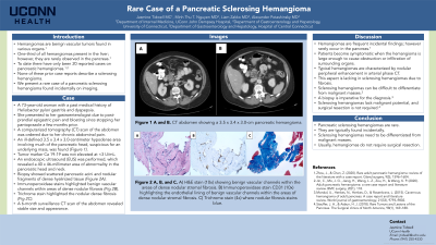
Case Description/Methods: A 73-year-old woman with a past medical history of Heliobacter pylori gastritis and dyspepsia previously on pantoprazole 40 mg daily presented to her gastroenterologist due to post-prandial epigastric pain and bloating since stopping her pantoprazole a few months prior. A computerized tomography (CT) scan of the abdomen was ordered due to her chronic abdominal pain. It revealed an incidental finding of an ill-defined 3.5 x 3.4 x 3.0 centimeter hypodense area involving much of the pancreatic head, suspicious for an underlying mass (Figure A and B). Tumor marker CA 19-19 was not elevated at < 3 U/mL.
An endoscopic ultrasound (EUS) was performed, which revealed a 40 x 46 millimeter area of abnormality in the pancreatic head and neck. An EUS biopsy was obtained which showed scattered pancreatic acini and nodular fragments of dense hyalinized tissue. A trichrome stain highlighted the nodular dense fibrosis and immunoperoxidase stains highlighted benign vascular channels within the areas of dense nodular fibrosis. The findings were consistent with a pancreatic sclerosing hemangioma. A 6-month surveillance CT scan of the abdomen revealed stable size and appearance. The patient’s dyspepsia resolved on pantoprazole therapy.
Discussion: We describe the rare finding of a pancreatic sclerosing hemangioma. Although hemangiomas are frequent incidental findings, they are rarely located in the pancreas. Most patients are asymptomatic unless the hemangioma grows large enough to cause obstruction or infiltration of surrounding organs. Typical hemangiomas are characterized by nodular peripheral enhancement in arterial phase on CT, which is lacking with sclerosing hemangiomas due to fibrosis. This makes it very difficult to differentiate from malignant masses, for which biopsy is imperative. Due to lack of malignant potential, it is important to prevent unnecessary surgical resection of sclerosing hemangiomas. It is essential that we be aware of pancreatic hemangiomas, especially if sclerosing, to ensure proper diagnostic work-up and surveillance.

Disclosures:
Jasmine Tidwell, MD1, Minh Thu T. Nguyen, MD1, Liam Zakko, MD2, Alexander Potashinksky, MD2. P2973 - Rare Case of a Pancreatic Sclerosing Hemangioma, ACG 2023 Annual Scientific Meeting Abstracts. Vancouver, BC, Canada: American College of Gastroenterology.
1UConn Health, Farmington, CT; 2Hospital of Central Connecticut, New Britain, CT
Introduction: Hemangiomas are benign vascular tumors found in various organs. One-third of all hemangiomas present in the liver; however, they are rarely observed in the pancreas. To date, there have only been 20 reported cases of pancreatic hemangiomas, and none described as sclerosing. We present a rare case of a sclerosing pancreatic hemangioma found incidentally on imaging.
Case Description/Methods: A 73-year-old woman with a past medical history of Heliobacter pylori gastritis and dyspepsia previously on pantoprazole 40 mg daily presented to her gastroenterologist due to post-prandial epigastric pain and bloating since stopping her pantoprazole a few months prior. A computerized tomography (CT) scan of the abdomen was ordered due to her chronic abdominal pain. It revealed an incidental finding of an ill-defined 3.5 x 3.4 x 3.0 centimeter hypodense area involving much of the pancreatic head, suspicious for an underlying mass (Figure A and B). Tumor marker CA 19-19 was not elevated at < 3 U/mL.
An endoscopic ultrasound (EUS) was performed, which revealed a 40 x 46 millimeter area of abnormality in the pancreatic head and neck. An EUS biopsy was obtained which showed scattered pancreatic acini and nodular fragments of dense hyalinized tissue. A trichrome stain highlighted the nodular dense fibrosis and immunoperoxidase stains highlighted benign vascular channels within the areas of dense nodular fibrosis. The findings were consistent with a pancreatic sclerosing hemangioma. A 6-month surveillance CT scan of the abdomen revealed stable size and appearance. The patient’s dyspepsia resolved on pantoprazole therapy.
Discussion: We describe the rare finding of a pancreatic sclerosing hemangioma. Although hemangiomas are frequent incidental findings, they are rarely located in the pancreas. Most patients are asymptomatic unless the hemangioma grows large enough to cause obstruction or infiltration of surrounding organs. Typical hemangiomas are characterized by nodular peripheral enhancement in arterial phase on CT, which is lacking with sclerosing hemangiomas due to fibrosis. This makes it very difficult to differentiate from malignant masses, for which biopsy is imperative. Due to lack of malignant potential, it is important to prevent unnecessary surgical resection of sclerosing hemangiomas. It is essential that we be aware of pancreatic hemangiomas, especially if sclerosing, to ensure proper diagnostic work-up and surveillance.

Figure: Figure A and B - CT scan of the abdomen evidencing a 3.5 x 3.4 x 3.0 centimeter hypodense area involving the pancreatic head.
Disclosures:
Jasmine Tidwell indicated no relevant financial relationships.
Minh Thu Nguyen indicated no relevant financial relationships.
Liam Zakko indicated no relevant financial relationships.
Alexander Potashinksky indicated no relevant financial relationships.
Jasmine Tidwell, MD1, Minh Thu T. Nguyen, MD1, Liam Zakko, MD2, Alexander Potashinksky, MD2. P2973 - Rare Case of a Pancreatic Sclerosing Hemangioma, ACG 2023 Annual Scientific Meeting Abstracts. Vancouver, BC, Canada: American College of Gastroenterology.
