Tuesday Poster Session
Category: Biliary/Pancreas
P2975 - Navigating the Ducts: Differentiating Between Caroli's Disease and Primary Sclerosing Cholangitis
Tuesday, October 24, 2023
10:30 AM - 4:00 PM PT
Location: Exhibit Hall


Anmol Mittal, MD
Rutgers New Jersey Medical School
Newark, NJ
Presenting Author(s)
Anmol Mittal, MD, Yazan Abboud, MD, Reza Hashemipour, MD, Kaveh Hajifathalian, MD, MPH
Rutgers New Jersey Medical School, Newark, NJ
Introduction: Caroli's disease is a rare genetic condition that is a difficult disease to diagnose. Compounded by the similarities with primary biliary cholangitis, the importance of highlighting patient presentations to guide more informed differentials is substantial. To our knowledge, there are few reports on Caroli's disease and even fewer remarking on hemobilia. Understanding this patient's presentation will assist in accurate diagnosis in future patients.
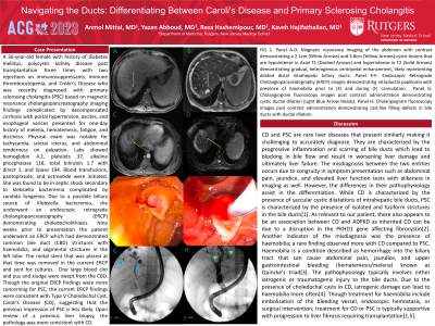
Case Description/Methods: A 36-year-old female with a history of polycystic kidney disease post-transplantation and Crohn's Disease who was recently diagnosed with primary sclerosing cholangitis (PSC) complicated by decompensated cirrhosis presented for one-day history of melena, hematemesis, fatigue, and dizziness. She was found to be in septic shock secondary to klebsiella bacteremia complicated by candida fungemia. She underwent an endoscopic retrograde cholangiopancreatography (ERCP) demonstrating choledocholithiasis. The metal stent that was placed at the ERCP performed nine weeks prior (when she was diagnosed with PSC) was removed during the current ERCP and sent for cultures. Though the original ERCP findings were more concerning for PSC, the current ERCP findings were consistent with Type V Choledochal Cyst, Caroli’s Disease (CD), suggesting that the previous impression of PSC is less likely. Upon review of a previous liver biopsy, the pathology was more consistent with CD.
Discussion: CD and PSC are rare liver diseases that present similarly making it challenging to accurately diagnose. They are characterized by the progressive inflammation and scarring of bile ducts which lead to blocked bile flow resulting in worsening liver damage and failure. While CD is characterized by saccular cystic dilatations of intrahepatic bile ducts, PSC is characterized by the presence of isolated and fusiform strictures in the bile ducts. As relevant to our patient, there also appears to be an association between CD and ADPKD as inherited CD can be due to a disruption in the PKHD1 gene affecting fibrocystin. Another indicator of the misdiagnosis was the presence of haemobilia; a rare finding observed more with CD compared to PSC. Due to the presence of choledochal cysts in CD, iatrogenic damage can lead to haemobilia more often. Though treatment for haemobilia includes embolization of the bleeding vessel, endoscopic hemostasis, or surgical intervention, treatment for CD or PSC is typically supportive with progression to liver fibrosis requiring transplantation.

Disclosures:
Anmol Mittal, MD, Yazan Abboud, MD, Reza Hashemipour, MD, Kaveh Hajifathalian, MD, MPH. P2975 - Navigating the Ducts: Differentiating Between Caroli's Disease and Primary Sclerosing Cholangitis, ACG 2023 Annual Scientific Meeting Abstracts. Vancouver, BC, Canada: American College of Gastroenterology.
Rutgers New Jersey Medical School, Newark, NJ
Introduction: Caroli's disease is a rare genetic condition that is a difficult disease to diagnose. Compounded by the similarities with primary biliary cholangitis, the importance of highlighting patient presentations to guide more informed differentials is substantial. To our knowledge, there are few reports on Caroli's disease and even fewer remarking on hemobilia. Understanding this patient's presentation will assist in accurate diagnosis in future patients.
Case Description/Methods: A 36-year-old female with a history of polycystic kidney disease post-transplantation and Crohn's Disease who was recently diagnosed with primary sclerosing cholangitis (PSC) complicated by decompensated cirrhosis presented for one-day history of melena, hematemesis, fatigue, and dizziness. She was found to be in septic shock secondary to klebsiella bacteremia complicated by candida fungemia. She underwent an endoscopic retrograde cholangiopancreatography (ERCP) demonstrating choledocholithiasis. The metal stent that was placed at the ERCP performed nine weeks prior (when she was diagnosed with PSC) was removed during the current ERCP and sent for cultures. Though the original ERCP findings were more concerning for PSC, the current ERCP findings were consistent with Type V Choledochal Cyst, Caroli’s Disease (CD), suggesting that the previous impression of PSC is less likely. Upon review of a previous liver biopsy, the pathology was more consistent with CD.
Discussion: CD and PSC are rare liver diseases that present similarly making it challenging to accurately diagnose. They are characterized by the progressive inflammation and scarring of bile ducts which lead to blocked bile flow resulting in worsening liver damage and failure. While CD is characterized by saccular cystic dilatations of intrahepatic bile ducts, PSC is characterized by the presence of isolated and fusiform strictures in the bile ducts. As relevant to our patient, there also appears to be an association between CD and ADPKD as inherited CD can be due to a disruption in the PKHD1 gene affecting fibrocystin. Another indicator of the misdiagnosis was the presence of haemobilia; a rare finding observed more with CD compared to PSC. Due to the presence of choledochal cysts in CD, iatrogenic damage can lead to haemobilia more often. Though treatment for haemobilia includes embolization of the bleeding vessel, endoscopic hemostasis, or surgical intervention, treatment for CD or PSC is typically supportive with progression to liver fibrosis requiring transplantation.

Figure: FIG 1. Panel A-D: Magnetic resonance imaging of the abdomen with contrast demonstrating a 2.1cm (White Arrows) and 3.8cm (Yellow Arrows) cystic lesions that are hypointense in Axial T1 (Dashed Arrows) and hyperintense in T2 (Solid Arrows) demonstrating gradual, heterogenous centripetal enhancement, likely representing dilated distal intrahepatic biliary ducts. Panel E-F: Endoscopic Retrograde Cholangiopancreatography (ERCP) images demonstrating intraductal papilloma with presence of haemobilia prior to (E) and during (F) cannulation. Panel G: Cholangiogram fluoroscopy images post contrast administration demonstrating cystic ductal dilation (Light Blue Arrow Heads). Panel H: Cholangiogram fluoroscopy images post contrast administration demonstrating cast-like filling defects in bile ducts with ductal dilation.
Disclosures:
Anmol Mittal indicated no relevant financial relationships.
Yazan Abboud indicated no relevant financial relationships.
Reza Hashemipour: Boston Scientific – Endoscopic Fellow Course Participant.
Kaveh Hajifathalian indicated no relevant financial relationships.
Anmol Mittal, MD, Yazan Abboud, MD, Reza Hashemipour, MD, Kaveh Hajifathalian, MD, MPH. P2975 - Navigating the Ducts: Differentiating Between Caroli's Disease and Primary Sclerosing Cholangitis, ACG 2023 Annual Scientific Meeting Abstracts. Vancouver, BC, Canada: American College of Gastroenterology.
