Tuesday Poster Session
Category: Biliary/Pancreas
P2983 - A Rare Case of Sarcoidosis in the Gallbladder
Tuesday, October 24, 2023
10:30 AM - 4:00 PM PT
Location: Exhibit Hall

Has Audio
- AM
Abdullah Memon, MD
University of Illinois Chicago
Chicago, IL
Presenting Author(s)
Abdullah Memon, MD1, Dexter E. Nwachukwu, DO, MS1, Adam E. Mikolajczyk, MD2, Miguel Alvarez, MD3, Grace Guzman, MD4, Matthew Niemeyer, MD1
1University of Illinois Chicago, Chicago, IL; 2University of Illinois Chicago, Highland, IN; 3University of Illinois at Chicago, Chicago, IL; 4University of Illinois at Chicago, College of Medicine, Chicago, IL
Introduction: Sarcoidosis is a multisystemic, noncaseating granulomatous disease that manifests primarily in the pulmonary and lymphoid system. It rarely affects the gastrointestinal system, with less than 4% of sarcoidosis cases having GI and hepatic involvement.
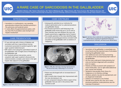
Case Description/Methods: We report a case of gallbladder sarcoidosis in a 37 year old male with history of diabetes and sarcoidosis with liver, lung and bone marrow involvement. He was initially admitted for right upper quadrant abdominal pain at an outside hospital three months before his visit at our institution. MRI there had noted a potential gallbladder lesion with T2 bright areas of thickening in the gallbladder wall, and his pain resolved without intervention. An outpatient right upper quadrant ultrasound after his discharge confirmed a 2.9 x 2.8 x 3.2 complex cystic lesion in the right hepatic lobe adjacent to the gallbladder. He subsequently was admitted at our institution for medical optimization prior to a planned procedure and gallbladder biopsy. EUS showed asymmetric wall thickening in the gallbladder body measuring up to 1.8 cm and an intact interface was seen between the mass and the hepatic parenchyma, suggesting a lack of invasion. Fine needle aspiration of the mass identified large and small noncaseating granulomas consistent with sarcoidosis of the gallbladder with no evidence of malignancy. Pt was discharged with an increased dose of prednisone, and MRI 3 months later noted clear improvement with the mass decreasing in size and the component inferior to the gallbladder resolved in the interval.
Discussion: Sarcoidosis of the gallbladder is exceedingly rare, as our literature review found only five reported cases of sarcoidosis involving the gallbladder alone. Four additional cases were found presenting sarcoidosis of the accompanying lymph nodes of the gallbladder. Out of these nine who all underwent cholecystectomy, only one had been diagnosed with sarcoidosis prior to surgery. As with our case, patients are likely to present with symptoms of acute cholecystitis, such as right-sided abdominal pain, nausea and vomiting. Patients may also be asymptomatic and are diagnosed incidentally. Additionally, symptoms can possibly mimic those of a malignancy or an infectious etiology, all which are part of the differential diagnosis. Evidence on treatment is very limited. Steroids were used for our patient, but in case reports previously published, cholecystectomy was the mainstay of treatment.

Disclosures:
Abdullah Memon, MD1, Dexter E. Nwachukwu, DO, MS1, Adam E. Mikolajczyk, MD2, Miguel Alvarez, MD3, Grace Guzman, MD4, Matthew Niemeyer, MD1. P2983 - A Rare Case of Sarcoidosis in the Gallbladder, ACG 2023 Annual Scientific Meeting Abstracts. Vancouver, BC, Canada: American College of Gastroenterology.
1University of Illinois Chicago, Chicago, IL; 2University of Illinois Chicago, Highland, IN; 3University of Illinois at Chicago, Chicago, IL; 4University of Illinois at Chicago, College of Medicine, Chicago, IL
Introduction: Sarcoidosis is a multisystemic, noncaseating granulomatous disease that manifests primarily in the pulmonary and lymphoid system. It rarely affects the gastrointestinal system, with less than 4% of sarcoidosis cases having GI and hepatic involvement.
Case Description/Methods: We report a case of gallbladder sarcoidosis in a 37 year old male with history of diabetes and sarcoidosis with liver, lung and bone marrow involvement. He was initially admitted for right upper quadrant abdominal pain at an outside hospital three months before his visit at our institution. MRI there had noted a potential gallbladder lesion with T2 bright areas of thickening in the gallbladder wall, and his pain resolved without intervention. An outpatient right upper quadrant ultrasound after his discharge confirmed a 2.9 x 2.8 x 3.2 complex cystic lesion in the right hepatic lobe adjacent to the gallbladder. He subsequently was admitted at our institution for medical optimization prior to a planned procedure and gallbladder biopsy. EUS showed asymmetric wall thickening in the gallbladder body measuring up to 1.8 cm and an intact interface was seen between the mass and the hepatic parenchyma, suggesting a lack of invasion. Fine needle aspiration of the mass identified large and small noncaseating granulomas consistent with sarcoidosis of the gallbladder with no evidence of malignancy. Pt was discharged with an increased dose of prednisone, and MRI 3 months later noted clear improvement with the mass decreasing in size and the component inferior to the gallbladder resolved in the interval.
Discussion: Sarcoidosis of the gallbladder is exceedingly rare, as our literature review found only five reported cases of sarcoidosis involving the gallbladder alone. Four additional cases were found presenting sarcoidosis of the accompanying lymph nodes of the gallbladder. Out of these nine who all underwent cholecystectomy, only one had been diagnosed with sarcoidosis prior to surgery. As with our case, patients are likely to present with symptoms of acute cholecystitis, such as right-sided abdominal pain, nausea and vomiting. Patients may also be asymptomatic and are diagnosed incidentally. Additionally, symptoms can possibly mimic those of a malignancy or an infectious etiology, all which are part of the differential diagnosis. Evidence on treatment is very limited. Steroids were used for our patient, but in case reports previously published, cholecystectomy was the mainstay of treatment.

Figure: Sections show scattered non-caseating granulomatous inflammation consisting of epithelioid histiocytes, lymphoplasmacytic infiltrates, and focal acute inflammation.
Disclosures:
Abdullah Memon indicated no relevant financial relationships.
Dexter Nwachukwu indicated no relevant financial relationships.
Adam Mikolajczyk indicated no relevant financial relationships.
Miguel Alvarez indicated no relevant financial relationships.
Grace Guzman indicated no relevant financial relationships.
Matthew Niemeyer indicated no relevant financial relationships.
Abdullah Memon, MD1, Dexter E. Nwachukwu, DO, MS1, Adam E. Mikolajczyk, MD2, Miguel Alvarez, MD3, Grace Guzman, MD4, Matthew Niemeyer, MD1. P2983 - A Rare Case of Sarcoidosis in the Gallbladder, ACG 2023 Annual Scientific Meeting Abstracts. Vancouver, BC, Canada: American College of Gastroenterology.
