Tuesday Poster Session
Category: Liver
P3986 - Hepatocellular Carcinoma Presenting as an Infiltrative Gallbladder Mass, a Rare Entity: Case Report and Literature Review
Tuesday, October 24, 2023
10:30 AM - 4:00 PM PT
Location: Exhibit Hall

Has Audio

Camille Lupianez-Merly, MD
Temple University Hospital
Philadelphia, PA
Presenting Author(s)
Camille Lupianez, MD, Jason Castellanos, MD, Kimberly Forde, MD, PhD, MHS
Temple University Hospital, Philadelphia, PA
Introduction: Hepatocellular carcinoma (HCC) is a primary liver malignancy generally characterized by intra-hepatic spread, with extrahepatic spread typically to lung, lymph nodes and bone. HCC originating from the gallbladder is an extremely rare entity. We report a case of a gallbladder mass infiltrating into the liver, initially thought to be a primary gallbladder carcinoma.
Case Description/Methods: 73-year-old female with hepatitis C virus s/p treatment and sustained virologic response, presented with chronic abdominal pain. Extensive workup including various imaging modalities, showed no significant findings except for a small portal venous thrombus (PVT) detected on MRA abdomen. 6 months later, she was diagnosed with COVID-19 and a pulmonary embolism. Continued abdominal pain prompted further evaluation with an abdominal US to evaluate for PVT extension revealing a large intraluminal gallbladder mass, with normal appearing liver. Follow-up CT showed a gallbladder mass, suspicious for primary malignancy, cirrhotic liver morphology without lesions. MRI confirmed a 4.6 cm gallbladder mass extending into the liver, suggestive of gallbladder malignancy or cholangiocarcinoma extending to the gallbladder. Tumor markers: AFP, Ca19-9, CEA were within normal range. Percutaneous biopsy of the gallbladder mass confirmed HCC. The patient was initiated on durvalumab/tremelimumab and underwent surgical resection.
Discussion: HCC comprises 80% of liver cancers. Metastasis to the gallbladder is rare, occurring in 2.8%-5.8% of all HCC cases, as it requires destruction of the gallbladder's muscular layer or collagen fibers to infiltrate the liver. In contrast, primary gallbladder carcinoma easily invades the liver due to the absence of peritoneum reflection. Our patient presented with a gallbladder mass infiltrating into the liver, a diagnostic dilemma, since this clinical scenario is more common for primary gallbladder cancer, than for HCC. Our patient had a small PVT, potentially indicating vascular migration via the portal system and cholecystic veins. Only a few cases of HCC presenting as a gallbladder mass have been reported in the literature, including our case. Pathophysiology and treatment for this condition lack consensus and prognosis for this aggressive tumor is generally poor. A multidisciplinary approach involving surgery and adjuvant chemotherapy may improve outcomes. Further research is needed to understand its behavior and establish treatment guidelines.

Disclosures:
Camille Lupianez, MD, Jason Castellanos, MD, Kimberly Forde, MD, PhD, MHS. P3986 - Hepatocellular Carcinoma Presenting as an Infiltrative Gallbladder Mass, a Rare Entity: Case Report and Literature Review, ACG 2023 Annual Scientific Meeting Abstracts. Vancouver, BC, Canada: American College of Gastroenterology.
Temple University Hospital, Philadelphia, PA
Introduction: Hepatocellular carcinoma (HCC) is a primary liver malignancy generally characterized by intra-hepatic spread, with extrahepatic spread typically to lung, lymph nodes and bone. HCC originating from the gallbladder is an extremely rare entity. We report a case of a gallbladder mass infiltrating into the liver, initially thought to be a primary gallbladder carcinoma.
Case Description/Methods: 73-year-old female with hepatitis C virus s/p treatment and sustained virologic response, presented with chronic abdominal pain. Extensive workup including various imaging modalities, showed no significant findings except for a small portal venous thrombus (PVT) detected on MRA abdomen. 6 months later, she was diagnosed with COVID-19 and a pulmonary embolism. Continued abdominal pain prompted further evaluation with an abdominal US to evaluate for PVT extension revealing a large intraluminal gallbladder mass, with normal appearing liver. Follow-up CT showed a gallbladder mass, suspicious for primary malignancy, cirrhotic liver morphology without lesions. MRI confirmed a 4.6 cm gallbladder mass extending into the liver, suggestive of gallbladder malignancy or cholangiocarcinoma extending to the gallbladder. Tumor markers: AFP, Ca19-9, CEA were within normal range. Percutaneous biopsy of the gallbladder mass confirmed HCC. The patient was initiated on durvalumab/tremelimumab and underwent surgical resection.
Discussion: HCC comprises 80% of liver cancers. Metastasis to the gallbladder is rare, occurring in 2.8%-5.8% of all HCC cases, as it requires destruction of the gallbladder's muscular layer or collagen fibers to infiltrate the liver. In contrast, primary gallbladder carcinoma easily invades the liver due to the absence of peritoneum reflection. Our patient presented with a gallbladder mass infiltrating into the liver, a diagnostic dilemma, since this clinical scenario is more common for primary gallbladder cancer, than for HCC. Our patient had a small PVT, potentially indicating vascular migration via the portal system and cholecystic veins. Only a few cases of HCC presenting as a gallbladder mass have been reported in the literature, including our case. Pathophysiology and treatment for this condition lack consensus and prognosis for this aggressive tumor is generally poor. A multidisciplinary approach involving surgery and adjuvant chemotherapy may improve outcomes. Further research is needed to understand its behavior and establish treatment guidelines.

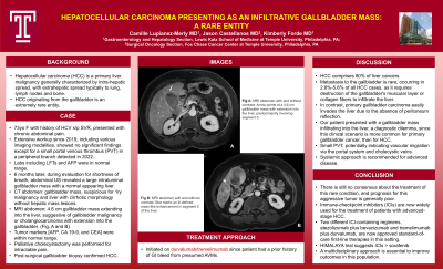
Figure: A. MRI abdomen with and without contrast: Arrow points at a 4.6 cm gallbladder mass with extension into the liver, predominantly involving segment 5.
B. MRI abdomen with and without contrast: Star marks an ill-defined mass-like enhancement in segment 5 of the liver adjacent to the gallbladder.
C. Segment 5/6 resection with en bloc cholecystectomy, posterior view. Arrow points at tumor invading gallbladder.
B. MRI abdomen with and without contrast: Star marks an ill-defined mass-like enhancement in segment 5 of the liver adjacent to the gallbladder.
C. Segment 5/6 resection with en bloc cholecystectomy, posterior view. Arrow points at tumor invading gallbladder.
Disclosures:
Camille Lupianez indicated no relevant financial relationships.
Jason Castellanos indicated no relevant financial relationships.
Kimberly Forde indicated no relevant financial relationships.
Camille Lupianez, MD, Jason Castellanos, MD, Kimberly Forde, MD, PhD, MHS. P3986 - Hepatocellular Carcinoma Presenting as an Infiltrative Gallbladder Mass, a Rare Entity: Case Report and Literature Review, ACG 2023 Annual Scientific Meeting Abstracts. Vancouver, BC, Canada: American College of Gastroenterology.
