Tuesday Poster Session
Category: Biliary/Pancreas
P2991 - Pancreatic Pseudocyst: A Presentation of Post-Acute Pancreatitis Complications: Summary and Recommendations in Management
Tuesday, October 24, 2023
10:30 AM - 4:00 PM PT
Location: Exhibit Hall

Has Audio
- EN
Emilio R. Narvaez, DO
Baylor Scott & White
Temple, TX
Presenting Author(s)
Emilio R. Narvaez, DO1, Christopher Chum, DO2, John Trillo, MD2, Nicholas Narvaez, 3, Stefan Friemel, MD4, Cory Robert Stephenson, DO4
1Baylor Scott & White, Temple, TX; 2NYCHHC South Brooklyn Health, Brooklyn, NY; 3FIU, Miami, FL; 4Baylor Scott & White Health, Temple, TX
Introduction: As our western society continues to indulge in an ever-increasing prevalence of high caloric diet, binge drinking and alcohol consumption, acute pancreatitis rates continue to rise. 20 to 40% of these acute pancreatitis cases progress toward pseudocyst formation and only 10 percent of these develop fistula formation, external or internal (2). This case demonstrates a rare complication of subacute pancreatitis, an internal fistula formation adjoining 2 hollow organs resulting in a pancreaticoduodenocolonic fistula with exceptional imaging.
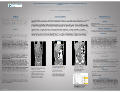
Case Description/Methods: The patient is a 30 y/o black male who presented 4 weeks after an initial episode of acute pancreatitis from an outside hospital in Atlanta, GA. induced by an episode of binge drinking. He denoted abdominal pain in the epigastrium, fevers, nausea, vomiting, and stools with mucus. He was having these symptoms for approximately 72 hours prior to presentation. Risk Factors included history of recurrent pancreatitis and heavy alcohol use.
Initial management was to begin broad spectrum antibiotic coverage with Zosyn and send the patient to interventional radiology after initial CT of the abdomen and pelvis showed a large pseudocyst causing mass effect on the liver, small bowel, stomach, colon and urinary bladder (figure 1). The cultures from this fluid grew out a pan sensitive E. Coli and his antibiotics were subsequently changed to ciprofloxacin (figure 3).
The patient subsequently complained of increased output from his pigtail catheter after having bowel movements. This raised the clinical suspicion of a fistula formation leading to the ultimate diagnosis of a pancreaticoduodenocolonic fistula a follow-up CT abdomen and pelvis imaging (figure 2). General surgery was involved and planned for laparoscopic debridement. The patient responded well, and surgical clips are noted at the sites of the duodenum and transverse colon on follow-up imaging (figure 4). There was no reason presentation of a pseudocyst formation post surgically.
Discussion: Familiarity and recognition of complications of pancreatitis at 4 weeks’ time is an important knowledge base for medical practice. Pancreatic pseudocysts formation is prevalent in 20 to 40% of presenting cases with a majority of these not complicated by infection and conservative management. This case demonstrated a rare complication of acute pancreatitis resulting in a pancreaticoduodenocolonic fistula formation.

Disclosures:
Emilio R. Narvaez, DO1, Christopher Chum, DO2, John Trillo, MD2, Nicholas Narvaez, 3, Stefan Friemel, MD4, Cory Robert Stephenson, DO4. P2991 - Pancreatic Pseudocyst: A Presentation of Post-Acute Pancreatitis Complications: Summary and Recommendations in Management, ACG 2023 Annual Scientific Meeting Abstracts. Vancouver, BC, Canada: American College of Gastroenterology.
1Baylor Scott & White, Temple, TX; 2NYCHHC South Brooklyn Health, Brooklyn, NY; 3FIU, Miami, FL; 4Baylor Scott & White Health, Temple, TX
Introduction: As our western society continues to indulge in an ever-increasing prevalence of high caloric diet, binge drinking and alcohol consumption, acute pancreatitis rates continue to rise. 20 to 40% of these acute pancreatitis cases progress toward pseudocyst formation and only 10 percent of these develop fistula formation, external or internal (2). This case demonstrates a rare complication of subacute pancreatitis, an internal fistula formation adjoining 2 hollow organs resulting in a pancreaticoduodenocolonic fistula with exceptional imaging.
Case Description/Methods: The patient is a 30 y/o black male who presented 4 weeks after an initial episode of acute pancreatitis from an outside hospital in Atlanta, GA. induced by an episode of binge drinking. He denoted abdominal pain in the epigastrium, fevers, nausea, vomiting, and stools with mucus. He was having these symptoms for approximately 72 hours prior to presentation. Risk Factors included history of recurrent pancreatitis and heavy alcohol use.
Initial management was to begin broad spectrum antibiotic coverage with Zosyn and send the patient to interventional radiology after initial CT of the abdomen and pelvis showed a large pseudocyst causing mass effect on the liver, small bowel, stomach, colon and urinary bladder (figure 1). The cultures from this fluid grew out a pan sensitive E. Coli and his antibiotics were subsequently changed to ciprofloxacin (figure 3).
The patient subsequently complained of increased output from his pigtail catheter after having bowel movements. This raised the clinical suspicion of a fistula formation leading to the ultimate diagnosis of a pancreaticoduodenocolonic fistula a follow-up CT abdomen and pelvis imaging (figure 2). General surgery was involved and planned for laparoscopic debridement. The patient responded well, and surgical clips are noted at the sites of the duodenum and transverse colon on follow-up imaging (figure 4). There was no reason presentation of a pseudocyst formation post surgically.
Discussion: Familiarity and recognition of complications of pancreatitis at 4 weeks’ time is an important knowledge base for medical practice. Pancreatic pseudocysts formation is prevalent in 20 to 40% of presenting cases with a majority of these not complicated by infection and conservative management. This case demonstrated a rare complication of acute pancreatitis resulting in a pancreaticoduodenocolonic fistula formation.

Figure: Figure 2: This is the pancreatic pseudocyst (PC) after placement of a pigtail catheter drain (D) you can see some resolution of the mass-effect and an air bubble inside of the pseudocyst. After placement the patient began to have improvement in compressive symptoms and tolerated progression of his diet. However, given his complaints of increased output after bowel movements a concern of pancreaticoduodenocolonic fistula is evident and noted by the radiologist. After this time general surgery was consulted as medical management with antibiotics without source control was unlikely to resolve the patient’s acute issue.
Disclosures:
Emilio Narvaez indicated no relevant financial relationships.
Christopher Chum indicated no relevant financial relationships.
John Trillo indicated no relevant financial relationships.
Nicholas Narvaez indicated no relevant financial relationships.
Stefan Friemel indicated no relevant financial relationships.
Cory Robert Stephenson indicated no relevant financial relationships.
Emilio R. Narvaez, DO1, Christopher Chum, DO2, John Trillo, MD2, Nicholas Narvaez, 3, Stefan Friemel, MD4, Cory Robert Stephenson, DO4. P2991 - Pancreatic Pseudocyst: A Presentation of Post-Acute Pancreatitis Complications: Summary and Recommendations in Management, ACG 2023 Annual Scientific Meeting Abstracts. Vancouver, BC, Canada: American College of Gastroenterology.
