Tuesday Poster Session
Category: Liver
P3996 - Giant Tumor Masquerading as Massive Mucinous Ascites
Tuesday, October 24, 2023
10:30 AM - 4:00 PM PT
Location: Exhibit Hall

Has Audio
- KM
Karthik Mathialagan, MD
Yale New Haven Health/Bridgeport Hospital
Bridgeport, CT
Presenting Author(s)
Karthik Mathialagan, MD1, Samdish Sethi, MD1, Prince Addo. Ameyaw, MD2, Prachi Pednekar, MD3
1Yale New Haven Health/Bridgeport Hospital, Bridgeport, CT; 2Bridgeport Hospital/Yale New Haven Health, Bridgeport, CT; 3Yale New Haven Hospital, New Haven, CT
Introduction: Ascites is the pathologic accumulation of fluid within the peritoneal cavity, with liver cirrhosis and portal hypertension accounting for 70% followed by malignancy in 10% of cases. In rare instances, ovarian lesions can cause ascites. We present a case of an ovarian mucinous tumor that ruptured prematurely leading to massive mucinous ascites resulting in a diagnostic dilemma.
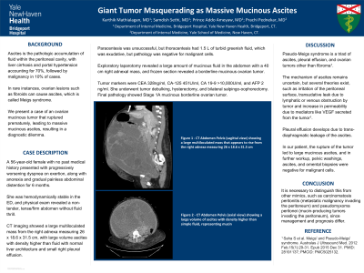
Case Description/Methods: A 56-year-old female with no past medical history presented with progressively worsening dyspnea on exertion along with anorexia and gradual painless abdominal distention for 6 months. In the ED, she was hemodynamically stable and physical exam revealed a nontender, tense/firm abdomen without fluid thrill. Initial blood work revealed mild microcytic anemia with normal liver and renal function. CT chest, abdomen/pelvis showed a large multiloculated mass from the right adnexa measuring 26 x 18.6 x 31.5 cm, surrounded by large volume ascites with density higher than fluid with normal liver architecture and small right pleural effusion. Paracentesis was unsuccessful but thoracentesis had 1.5 L of turbid greenish fluid which was exudative, but pathology was negative for malignant cells. EGD and colonoscopy ruled out GI tumors. Eventually, she underwent an exploratory laparotomy which revealed a large amount of mucinous fluid in the abdomen with a 40 cm right adnexal mass and frozen section revealed a borderline mucinous ovarian tumor. Tumor markers were CEA 328ng/ml, CA-125 451U/ml, CA 19-9 >10,000U/ml and AFP of 2 ng/ml. She underwent tumor debulking, hysterectomy and bilateral salpingo-oophorectomy. Final pathology showed Stage 1A mucinous borderline ovarian tumor.
Discussion: Pseudo-Meigs syndrome is a triad of ascites, pleural effusion and ovarian tumors other than fibroma. The mechanism of ascites remains uncertain, but several theories exist, such as irritation of peritoneal surface, transudative leak due to lymphatic or venous obstruction by tumor, increase in permeability due to mediators secreted from tumor. Pleural effusion develops due to trans-diaphragmatic leakage of the ascites. In our patient, rupture of the tumor led to large mucinous ascites. It is necessary to distinguish this from other mimics, such as carcinomatosis peritonitis (metastatic malignancy invading the peritoneum) and pseudomyxoma peritonei (mucin producing tumors invading the peritoneum) since management and prognosis differ. In our patient, pelvic washings, ascites and omental biopsies were negative for malignant cells.

Disclosures:
Karthik Mathialagan, MD1, Samdish Sethi, MD1, Prince Addo. Ameyaw, MD2, Prachi Pednekar, MD3. P3996 - Giant Tumor Masquerading as Massive Mucinous Ascites, ACG 2023 Annual Scientific Meeting Abstracts. Vancouver, BC, Canada: American College of Gastroenterology.
1Yale New Haven Health/Bridgeport Hospital, Bridgeport, CT; 2Bridgeport Hospital/Yale New Haven Health, Bridgeport, CT; 3Yale New Haven Hospital, New Haven, CT
Introduction: Ascites is the pathologic accumulation of fluid within the peritoneal cavity, with liver cirrhosis and portal hypertension accounting for 70% followed by malignancy in 10% of cases. In rare instances, ovarian lesions can cause ascites. We present a case of an ovarian mucinous tumor that ruptured prematurely leading to massive mucinous ascites resulting in a diagnostic dilemma.
Case Description/Methods: A 56-year-old female with no past medical history presented with progressively worsening dyspnea on exertion along with anorexia and gradual painless abdominal distention for 6 months. In the ED, she was hemodynamically stable and physical exam revealed a nontender, tense/firm abdomen without fluid thrill. Initial blood work revealed mild microcytic anemia with normal liver and renal function. CT chest, abdomen/pelvis showed a large multiloculated mass from the right adnexa measuring 26 x 18.6 x 31.5 cm, surrounded by large volume ascites with density higher than fluid with normal liver architecture and small right pleural effusion. Paracentesis was unsuccessful but thoracentesis had 1.5 L of turbid greenish fluid which was exudative, but pathology was negative for malignant cells. EGD and colonoscopy ruled out GI tumors. Eventually, she underwent an exploratory laparotomy which revealed a large amount of mucinous fluid in the abdomen with a 40 cm right adnexal mass and frozen section revealed a borderline mucinous ovarian tumor. Tumor markers were CEA 328ng/ml, CA-125 451U/ml, CA 19-9 >10,000U/ml and AFP of 2 ng/ml. She underwent tumor debulking, hysterectomy and bilateral salpingo-oophorectomy. Final pathology showed Stage 1A mucinous borderline ovarian tumor.
Discussion: Pseudo-Meigs syndrome is a triad of ascites, pleural effusion and ovarian tumors other than fibroma. The mechanism of ascites remains uncertain, but several theories exist, such as irritation of peritoneal surface, transudative leak due to lymphatic or venous obstruction by tumor, increase in permeability due to mediators secreted from tumor. Pleural effusion develops due to trans-diaphragmatic leakage of the ascites. In our patient, rupture of the tumor led to large mucinous ascites. It is necessary to distinguish this from other mimics, such as carcinomatosis peritonitis (metastatic malignancy invading the peritoneum) and pseudomyxoma peritonei (mucin producing tumors invading the peritoneum) since management and prognosis differ. In our patient, pelvic washings, ascites and omental biopsies were negative for malignant cells.

Figure: A - CT Abdomen Pelvis (sagittal view) showing a large multiloculated mass that appears to rise from the right adnexa measuring 26 x 18.6 x 31.5 cm.
B - CT Abdomen Pelvis (axial view) showing a large volume of ascites with density higher than simple fluid, representing mucin.
C - CT Chest showing a small right-sided pleural effusion with adjacent atelectasis.
B - CT Abdomen Pelvis (axial view) showing a large volume of ascites with density higher than simple fluid, representing mucin.
C - CT Chest showing a small right-sided pleural effusion with adjacent atelectasis.
Disclosures:
Karthik Mathialagan indicated no relevant financial relationships.
Samdish Sethi indicated no relevant financial relationships.
Prince Ameyaw indicated no relevant financial relationships.
Prachi Pednekar indicated no relevant financial relationships.
Karthik Mathialagan, MD1, Samdish Sethi, MD1, Prince Addo. Ameyaw, MD2, Prachi Pednekar, MD3. P3996 - Giant Tumor Masquerading as Massive Mucinous Ascites, ACG 2023 Annual Scientific Meeting Abstracts. Vancouver, BC, Canada: American College of Gastroenterology.
