Tuesday Poster Session
Category: Colon
P3048 - Amicable Amebiasis: A Case of Asymptomatic Intestinal Entamoeba histolytica
Tuesday, October 24, 2023
10:30 AM - 4:00 PM PT
Location: Exhibit Hall

Has Audio

Cherry W. Huang, DO
Walter Reed National Military Medical Center
Bethesda, MD
Presenting Author(s)
Cherry W. Huang, DO, Andrew Mertz, MD, Jessica Nicole, MD, Patrick Young, MD, FACG
Walter Reed National Military Medical Center, Bethesda, MD
Introduction: Amebiasis, caused by the protozoan Entamoeba histolytica, is the leading cause of infectious diarrhea worldwide. Areas with highest rates of infection include India, parts of Central and South America, Mexico, and Africa. Worldwide, amebiasis affects more than 50 million people with approximately 100,000 deaths annually. Though amebiasis is considered endemic worldwide, the prevalence of infections in the United States is relatively low, estimated to be about 1-4%. Interestingly, despite its global impact, E. histolytica infections are largely asymptomatic, with only 10-20% of those infected develop symptoms. Typically, intestinal amebiasis is diagnosed via stool microscopy, stool antigen detection, or stool polymerase chain reaction. Herein, we present a case of patient intestinal amebiasis diagnosed via routine screening colonoscopy.
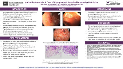
Case Description/Methods: The patient is a 56-year-old healthy male who was referred to the gastroenterology clinic for an index colonoscopy. He had no personal or family history of colorectal cancer. He denied any constitutional symptoms like fevers, chills, weight loss or malaise. He also denied any gastrointestinal symptoms including decreased appetite, nausea, vomiting, abdominal pain, diarrhea, constipation, and bloody bowel movements. Upon further questioning, the patient states that he often travels for work. Two months prior to his colonoscopy, he was in Japan for about a month. Within the past year, he has traveled to Australia and Norway, each visit staying for approximately a month. To his recollection, he usually drinks bottled water and eats cooked food, except for raw fish and sushi in Japan. His colonoscopy showed multifocal cecal ulcerations with surrounding exudate and friability. Biopsy samples taken from the cecum showed active colitis with organisms consistent with E. histolytica. He was treated with a 7-day course of paromomycin.
Discussion: This case highlights the importance of maintaining a broad differential diagnosis for colonic ulcerations and obtaining a good social history, including travel history for all patients. The endoscopic and histopathologic appearance of the mucosal lesions and their biopsies in this case are hallmarks of the disease but clinicians in the US may not consider it due to low prevalence. It is important to correctly identify and treat amebiasis to prevent the risk of worsening disease and decrease the risk of spread to family members.

Disclosures:
Cherry W. Huang, DO, Andrew Mertz, MD, Jessica Nicole, MD, Patrick Young, MD, FACG. P3048 - Amicable Amebiasis: A Case of Asymptomatic Intestinal Entamoeba histolytica, ACG 2023 Annual Scientific Meeting Abstracts. Vancouver, BC, Canada: American College of Gastroenterology.
Walter Reed National Military Medical Center, Bethesda, MD
Introduction: Amebiasis, caused by the protozoan Entamoeba histolytica, is the leading cause of infectious diarrhea worldwide. Areas with highest rates of infection include India, parts of Central and South America, Mexico, and Africa. Worldwide, amebiasis affects more than 50 million people with approximately 100,000 deaths annually. Though amebiasis is considered endemic worldwide, the prevalence of infections in the United States is relatively low, estimated to be about 1-4%. Interestingly, despite its global impact, E. histolytica infections are largely asymptomatic, with only 10-20% of those infected develop symptoms. Typically, intestinal amebiasis is diagnosed via stool microscopy, stool antigen detection, or stool polymerase chain reaction. Herein, we present a case of patient intestinal amebiasis diagnosed via routine screening colonoscopy.
Case Description/Methods: The patient is a 56-year-old healthy male who was referred to the gastroenterology clinic for an index colonoscopy. He had no personal or family history of colorectal cancer. He denied any constitutional symptoms like fevers, chills, weight loss or malaise. He also denied any gastrointestinal symptoms including decreased appetite, nausea, vomiting, abdominal pain, diarrhea, constipation, and bloody bowel movements. Upon further questioning, the patient states that he often travels for work. Two months prior to his colonoscopy, he was in Japan for about a month. Within the past year, he has traveled to Australia and Norway, each visit staying for approximately a month. To his recollection, he usually drinks bottled water and eats cooked food, except for raw fish and sushi in Japan. His colonoscopy showed multifocal cecal ulcerations with surrounding exudate and friability. Biopsy samples taken from the cecum showed active colitis with organisms consistent with E. histolytica. He was treated with a 7-day course of paromomycin.
Discussion: This case highlights the importance of maintaining a broad differential diagnosis for colonic ulcerations and obtaining a good social history, including travel history for all patients. The endoscopic and histopathologic appearance of the mucosal lesions and their biopsies in this case are hallmarks of the disease but clinicians in the US may not consider it due to low prevalence. It is important to correctly identify and treat amebiasis to prevent the risk of worsening disease and decrease the risk of spread to family members.

Figure: Amebiasis on Colonoscopy: Multifocal cecal ulcerations
Disclosures:
Cherry Huang indicated no relevant financial relationships.
Andrew Mertz indicated no relevant financial relationships.
Jessica Nicole indicated no relevant financial relationships.
Patrick Young indicated no relevant financial relationships.
Cherry W. Huang, DO, Andrew Mertz, MD, Jessica Nicole, MD, Patrick Young, MD, FACG. P3048 - Amicable Amebiasis: A Case of Asymptomatic Intestinal Entamoeba histolytica, ACG 2023 Annual Scientific Meeting Abstracts. Vancouver, BC, Canada: American College of Gastroenterology.
