Tuesday Poster Session
Category: Colon
P3052 - Sevelamer Induced Gastrointestinal Injury
Tuesday, October 24, 2023
10:30 AM - 4:00 PM PT
Location: Exhibit Hall

Has Audio
- SB
Sonya Bhaskar, MD
University of South Florida
Tampa, Florida
Presenting Author(s)
Sonya Bhaskar, MD1, Cesar Taborda-Vidarte, MD2, Wojciech Blonski, MD, PhD3
1University of South Florida, Tampa, FL; 2University of South Florida/James A. Haley VA Hospital, Tampa, FL; 3James A. Haley Veteran's Hospital and USF Health, Tampa, FL
Introduction: Sevelamer is a mainstay in the treatment of hyperphosphatemia in patients with chronic kidney disease which works by binding phosphate in the intestinal lumen to limit the absorption and decrease phosphate concentrations. Here, we describe two cases of sevelamer deposition causing adverse gastrointestinal effects.
Case Description/Methods: Case 1: A 62-year-old female with end-stage renal disease (ESRD) on hemodialysis (HD), and atrial fibrillation reported intermittent, cramping, right sided abdominal pain for two days with no association to oral intake. Patient was hemodynamically stable and labs were at her baseline. A CT Abdomen/Pelvis showed cecal wall thickening. Colonoscopy was performed and white-yellow exudate with surrounding erythema was seen in the distal ascending colon to the ileocecal valve. Given concerns for acute mesenteric ischemia, a lactic acid and CT angiography were obtained. Lactic acid was normal and CTA demonstrated the previous findings of colitis. Her symptoms resolved and she was advised to have a repeat colonoscopy in 2 months. Pathology resulted and showed necrotic tissue and fibrinopurulent exudate, compatible with ulcer debris with admixed sevelamer resin crystals. Sevelamer was discontinued.
Case 2: A 62 year old male with ESRD on HD and heart failure presented with five days of painless hematochezia with no associated symptoms. His hemoglobin had down trended from his baseline of 9-10 to 8.6 and he was hemodynamically stable. Colonoscopy showed areas of inflammation characterized by edema, erythema and ulcerations in the transverse and ascending colon, diverticulosis and hemorrhoids. Patient was discharged with resolution of symptoms. Biopsies showed microscopic/ischemic injury patterns with fish scale crystalline material. Sevelamer was discontinued.
Discussion: Given the right sided mucosal changes seen in the first case, the initial concern was to rule out etiologies of acute mesenteric ischemia such as arterial embolism, thromboembolism or vasculitis with a CTA given the risk factors of atrial fibrillation and ESRD. In our second case, the initial differential included ischemia given his risk factors of ESRD and heart failure. Our cases broaden the differential in this patient population and add to a growing number of cases of Sevelamer-related mucosal injury which include colitis, impaction, ulcerations, necrosis, and perforations throughout the gastrointestinal tract. The mechanism of action remains unclear, and treatment involves avoidance of the drug.

Disclosures:
Sonya Bhaskar, MD1, Cesar Taborda-Vidarte, MD2, Wojciech Blonski, MD, PhD3. P3052 - Sevelamer Induced Gastrointestinal Injury, ACG 2023 Annual Scientific Meeting Abstracts. Vancouver, BC, Canada: American College of Gastroenterology.
1University of South Florida, Tampa, FL; 2University of South Florida/James A. Haley VA Hospital, Tampa, FL; 3James A. Haley Veteran's Hospital and USF Health, Tampa, FL
Introduction: Sevelamer is a mainstay in the treatment of hyperphosphatemia in patients with chronic kidney disease which works by binding phosphate in the intestinal lumen to limit the absorption and decrease phosphate concentrations. Here, we describe two cases of sevelamer deposition causing adverse gastrointestinal effects.
Case Description/Methods: Case 1: A 62-year-old female with end-stage renal disease (ESRD) on hemodialysis (HD), and atrial fibrillation reported intermittent, cramping, right sided abdominal pain for two days with no association to oral intake. Patient was hemodynamically stable and labs were at her baseline. A CT Abdomen/Pelvis showed cecal wall thickening. Colonoscopy was performed and white-yellow exudate with surrounding erythema was seen in the distal ascending colon to the ileocecal valve. Given concerns for acute mesenteric ischemia, a lactic acid and CT angiography were obtained. Lactic acid was normal and CTA demonstrated the previous findings of colitis. Her symptoms resolved and she was advised to have a repeat colonoscopy in 2 months. Pathology resulted and showed necrotic tissue and fibrinopurulent exudate, compatible with ulcer debris with admixed sevelamer resin crystals. Sevelamer was discontinued.
Case 2: A 62 year old male with ESRD on HD and heart failure presented with five days of painless hematochezia with no associated symptoms. His hemoglobin had down trended from his baseline of 9-10 to 8.6 and he was hemodynamically stable. Colonoscopy showed areas of inflammation characterized by edema, erythema and ulcerations in the transverse and ascending colon, diverticulosis and hemorrhoids. Patient was discharged with resolution of symptoms. Biopsies showed microscopic/ischemic injury patterns with fish scale crystalline material. Sevelamer was discontinued.
Discussion: Given the right sided mucosal changes seen in the first case, the initial concern was to rule out etiologies of acute mesenteric ischemia such as arterial embolism, thromboembolism or vasculitis with a CTA given the risk factors of atrial fibrillation and ESRD. In our second case, the initial differential included ischemia given his risk factors of ESRD and heart failure. Our cases broaden the differential in this patient population and add to a growing number of cases of Sevelamer-related mucosal injury which include colitis, impaction, ulcerations, necrosis, and perforations throughout the gastrointestinal tract. The mechanism of action remains unclear, and treatment involves avoidance of the drug.

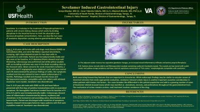
Figure: Case 1:
A: The adjacent viable mucosa has reparative glandular changes, an increased mixed inflammatory infiltrate and lamina propria fibroplasia.
B-D: Sections show necrotic debris and fibrinopurulent exudate containing scattered rhomboid crystals. The crystals are two toned with a pink center and yellow-orange periphery and have internal septations reminiscent of “fish scales,” morphologically compatible with sevelamer.
Disclosures:
Sonya Bhaskar indicated no relevant financial relationships.
Cesar Taborda-Vidarte indicated no relevant financial relationships.
Wojciech Blonski indicated no relevant financial relationships.
Sonya Bhaskar, MD1, Cesar Taborda-Vidarte, MD2, Wojciech Blonski, MD, PhD3. P3052 - Sevelamer Induced Gastrointestinal Injury, ACG 2023 Annual Scientific Meeting Abstracts. Vancouver, BC, Canada: American College of Gastroenterology.
