Tuesday Poster Session
Category: Colon
P3072 - Polypoid Arteriovenous Malformation of the Colon: An Extremely Rare Case
Tuesday, October 24, 2023
10:30 AM - 4:00 PM PT
Location: Exhibit Hall

Has Audio
- BS
Bishr Swar, MD
College of Medicine, University of Oklahoma Health Sciences Center
Oklahoma City, OK
Presenting Author(s)
Bishr Swar, MD1, Muhammad Taha Khan, MD2, Hashroop Gurm, MD3, Donald Kastens, MD4
1College of Medicine, University of Oklahoma Health Sciences Center, Oklahoma City, OK; 2Oklahoma University Health Sciences Center, Oklahoma City, OK; 3University of Oklahoma College of Medicine, Oklahoma City, OK; 4University of Oklahoma College of Medicine, Oklahoma CIty, OK
Introduction: Colonic angiodysplasia is characterized by dilated, thin-walled blood vessels which may present as GI bleeding and appear as flat, red, fern-like lesions on colonoscopy. We describe an extraordinarily rare case of an incidental finding of polypoid arteriovenous malformation (AVM) only diagnosed after histologic examination.
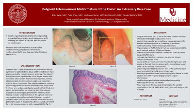
Case Description/Methods: We describe a 51-year-old male with a past medical history significant for polysubstance abuse and traumatic brain injury who presented for colorectal cancer screening. The patients family history was significant for a first degree relative with metastatic colon cancer. He denied hematochezia, melena, abdominal pain, or unintentional weight loss. Laboratory analysis was unremarkable. The patient underwent screening colonoscopy to the cecum with excellent bowel preparation. A 12 mm semi-pedunculated polyp was identified, lifted with saline, and removed by hot snare (Fig A-B). The area was clipped x3 to prevent post-polypectomy bleeding. Other findings included small diverticula in the sigmoid colon and non-bleeding internal hemorrhoids. Pathology revealed polypoid colonic mucosa with thickened muscularis mucosa, prominent vessels in mucosa and submucosa, and prominent fat in the mucosa consistent with a polypoid AVM (Fig C).
Discussion: The gastrointestinal tract is one of the more common locations where aberrant blood vessels can be found. Most of these vascular abnormalities are acquired later in life and can commonly present as GI bleeding or are found incidentally during routine endoscopic evaluation. Angiodysplasia or AVM of the GI tract are associated with ESRD, aortic stenosis, and von Willebrand disease. A review of the literature reveals15 reported cases of colonic polypoid AVM lesions. Histologically these lesions display characteristic dilated, tortuous submucosal veins. Colonic AVMs are most commonly found in the right colon and appear as flat, red, fern-like lesions with a central blood vessel giving rise to radiating ectatic vessels. GI bleeding related to these lesions tend to have chronic occult blood loss rather than brisk, overt hemorrhage. Bleeding is generally treated endoscopically with thermal or laser ablation and rarely requires angiographic or surgical interventions. Nonbleeding angiodysplasias incidentally discovered by endoscopy are usually not treated. Thus, we present this case that demonstrates the variable morphology of colonic AVMs which may even rarely masquerade as polyps.

Disclosures:
Bishr Swar, MD1, Muhammad Taha Khan, MD2, Hashroop Gurm, MD3, Donald Kastens, MD4. P3072 - Polypoid Arteriovenous Malformation of the Colon: An Extremely Rare Case, ACG 2023 Annual Scientific Meeting Abstracts. Vancouver, BC, Canada: American College of Gastroenterology.
1College of Medicine, University of Oklahoma Health Sciences Center, Oklahoma City, OK; 2Oklahoma University Health Sciences Center, Oklahoma City, OK; 3University of Oklahoma College of Medicine, Oklahoma City, OK; 4University of Oklahoma College of Medicine, Oklahoma CIty, OK
Introduction: Colonic angiodysplasia is characterized by dilated, thin-walled blood vessels which may present as GI bleeding and appear as flat, red, fern-like lesions on colonoscopy. We describe an extraordinarily rare case of an incidental finding of polypoid arteriovenous malformation (AVM) only diagnosed after histologic examination.
Case Description/Methods: We describe a 51-year-old male with a past medical history significant for polysubstance abuse and traumatic brain injury who presented for colorectal cancer screening. The patients family history was significant for a first degree relative with metastatic colon cancer. He denied hematochezia, melena, abdominal pain, or unintentional weight loss. Laboratory analysis was unremarkable. The patient underwent screening colonoscopy to the cecum with excellent bowel preparation. A 12 mm semi-pedunculated polyp was identified, lifted with saline, and removed by hot snare (Fig A-B). The area was clipped x3 to prevent post-polypectomy bleeding. Other findings included small diverticula in the sigmoid colon and non-bleeding internal hemorrhoids. Pathology revealed polypoid colonic mucosa with thickened muscularis mucosa, prominent vessels in mucosa and submucosa, and prominent fat in the mucosa consistent with a polypoid AVM (Fig C).
Discussion: The gastrointestinal tract is one of the more common locations where aberrant blood vessels can be found. Most of these vascular abnormalities are acquired later in life and can commonly present as GI bleeding or are found incidentally during routine endoscopic evaluation. Angiodysplasia or AVM of the GI tract are associated with ESRD, aortic stenosis, and von Willebrand disease. A review of the literature reveals15 reported cases of colonic polypoid AVM lesions. Histologically these lesions display characteristic dilated, tortuous submucosal veins. Colonic AVMs are most commonly found in the right colon and appear as flat, red, fern-like lesions with a central blood vessel giving rise to radiating ectatic vessels. GI bleeding related to these lesions tend to have chronic occult blood loss rather than brisk, overt hemorrhage. Bleeding is generally treated endoscopically with thermal or laser ablation and rarely requires angiographic or surgical interventions. Nonbleeding angiodysplasias incidentally discovered by endoscopy are usually not treated. Thus, we present this case that demonstrates the variable morphology of colonic AVMs which may even rarely masquerade as polyps.

Figure: Figure (A), Figure (B), Figure (C)
Disclosures:
Bishr Swar indicated no relevant financial relationships.
Muhammad Taha Khan indicated no relevant financial relationships.
Hashroop Gurm indicated no relevant financial relationships.
Donald Kastens indicated no relevant financial relationships.
Bishr Swar, MD1, Muhammad Taha Khan, MD2, Hashroop Gurm, MD3, Donald Kastens, MD4. P3072 - Polypoid Arteriovenous Malformation of the Colon: An Extremely Rare Case, ACG 2023 Annual Scientific Meeting Abstracts. Vancouver, BC, Canada: American College of Gastroenterology.
