Tuesday Poster Session
Category: Colon
P3078 - Colon Cancer: An Unexpected Cause of Dysphagia
Tuesday, October 24, 2023
10:30 AM - 4:00 PM PT
Location: Exhibit Hall

Has Audio

Kimberly Kucharski, MD
University of Utah
Presenting Author(s)
Award: Presidential Poster Award
Kim Kucharski, MD, Jessica Stout, DO
University of Utah, Salt Lake City, UT
Introduction: The colon can be partially resected and used to replace a section of the esophagus in patients with a variety of conditions including tracheoesophageal fistula, esophageal atresia, caustic ingestion, or after esophagectomy for esophageal cancer. This surgical procedure is known as colonic interposition. The development of malignancy in the colonic interposition graft is rare, with less than 20 cases reported in the literature. We present a case of a young female with adenocarcinoma of a colonic interposition in the esophagus.
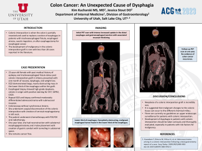
Case Description/Methods: A 23-year-old female patient with history of epilepsy and tracheoesophageal fistula status post colonic interposition graft in infancy presented with one month of nausea, dysphagia, and weight loss. At time of presentation, the patient’s only medication was oxcarbazepine. Esophagogastroduodenoscopy (EGD) was performed which demonstrated a large, nearly obstructing mass in the lower third of the esophagus within the graft. Initial esophageal biopsy showed high-grade dysplasia, colonic in origin with positive staining for CK7, SATB2, and CK20. Repeat EGD and biopsy shortly thereafter confirmed moderately differentiated adenocarcinoma with submucosal invasion. Further workup with a positron emission tomography (PET) scan revealed intense increased uptake in the distal esophagus and gastroesophageal junction with associated mucosal thickening. The PET scan was otherwise unremarkable for metastatic disease. Colonoscopy was performed and without synchronous lesions. She underwent surgical disconnection of the colonic interposition with creation of cervical esophagostomy in left neck. Intraoperatively, the distal colon interposition was found to be densely adherent to mediastinal structures and liver, with invasion into the lateral wall of the inferior vena cava. The patient subsequently underwent chemotherapy with FOLFOX and radiotherapy. Follow-up imaging after treatment is pending.
Discussion: Neoplasia of a colonic interposition graft is incredibly rare. Some experts suspect that malignant changes to the colonic tissue can occur due to the different chemical milieu.1 There are currently no guidelines on upper endoscopic surveillance for patients with colonic interposition. Development of dysphagia in a patient with colonic interposition should always be taken seriously and thoroughly evaluated, especially in patients with risk factors for malignancy including history of colonic polyps, colitis, or family history of colon cancer.

Disclosures:
Kim Kucharski, MD, Jessica Stout, DO. P3078 - Colon Cancer: An Unexpected Cause of Dysphagia, ACG 2023 Annual Scientific Meeting Abstracts. Vancouver, BC, Canada: American College of Gastroenterology.
Kim Kucharski, MD, Jessica Stout, DO
University of Utah, Salt Lake City, UT
Introduction: The colon can be partially resected and used to replace a section of the esophagus in patients with a variety of conditions including tracheoesophageal fistula, esophageal atresia, caustic ingestion, or after esophagectomy for esophageal cancer. This surgical procedure is known as colonic interposition. The development of malignancy in the colonic interposition graft is rare, with less than 20 cases reported in the literature. We present a case of a young female with adenocarcinoma of a colonic interposition in the esophagus.
Case Description/Methods: A 23-year-old female patient with history of epilepsy and tracheoesophageal fistula status post colonic interposition graft in infancy presented with one month of nausea, dysphagia, and weight loss. At time of presentation, the patient’s only medication was oxcarbazepine. Esophagogastroduodenoscopy (EGD) was performed which demonstrated a large, nearly obstructing mass in the lower third of the esophagus within the graft. Initial esophageal biopsy showed high-grade dysplasia, colonic in origin with positive staining for CK7, SATB2, and CK20. Repeat EGD and biopsy shortly thereafter confirmed moderately differentiated adenocarcinoma with submucosal invasion. Further workup with a positron emission tomography (PET) scan revealed intense increased uptake in the distal esophagus and gastroesophageal junction with associated mucosal thickening. The PET scan was otherwise unremarkable for metastatic disease. Colonoscopy was performed and without synchronous lesions. She underwent surgical disconnection of the colonic interposition with creation of cervical esophagostomy in left neck. Intraoperatively, the distal colon interposition was found to be densely adherent to mediastinal structures and liver, with invasion into the lateral wall of the inferior vena cava. The patient subsequently underwent chemotherapy with FOLFOX and radiotherapy. Follow-up imaging after treatment is pending.
Discussion: Neoplasia of a colonic interposition graft is incredibly rare. Some experts suspect that malignant changes to the colonic tissue can occur due to the different chemical milieu.1 There are currently no guidelines on upper endoscopic surveillance for patients with colonic interposition. Development of dysphagia in a patient with colonic interposition should always be taken seriously and thoroughly evaluated, especially in patients with risk factors for malignancy including history of colonic polyps, colitis, or family history of colon cancer.

Figure: PET scan with intense increased uptake in the distal esophagus and gastroesophageal junction with associated mucosal thickening
Disclosures:
Kim Kucharski indicated no relevant financial relationships.
Jessica Stout indicated no relevant financial relationships.
Kim Kucharski, MD, Jessica Stout, DO. P3078 - Colon Cancer: An Unexpected Cause of Dysphagia, ACG 2023 Annual Scientific Meeting Abstracts. Vancouver, BC, Canada: American College of Gastroenterology.

