Tuesday Poster Session
Category: Colon
P3086 - Rectal Schwannoma: A Rare Gastrointestinal Tumor
Tuesday, October 24, 2023
10:30 AM - 4:00 PM PT
Location: Exhibit Hall

Has Audio

Arooj Mian, MD
AdventHealth
Orlando, FL
Presenting Author(s)
Arooj Mian, MD1, Gurdeep Singh, DO1, Abdul Mohammed, MD2, Ilan Aharoni, MD2
1AdventHealth, Orlando, FL; 2AdventHealth, Winter Park, FL
Introduction: Schwannomas are benign spindle cell tumors originating from Schwann cells, which form the neural sheath. Gastrointestinal (GI) schwannomas are rare, accounting for 3% of all GI mesenchymal tumors. When present, GI schwannomas most frequently occur in the stomach (83%) and small intestine (12%) and rarely involve the colon and rectum. We report a case of rectal schwannoma in a patient with type 2 neurofibromatosis.
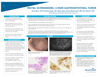
Case Description/Methods: A 36-year-old male with a history of type 2 neurofibromatosis, bilateral acoustic schwannomas, and cervical and lumbosacral spine schwannomas treated with CyberKnife and Bevacizumab chemotherapy presented with change in bowel habits, fecal urgency, and fecal incontinence of several months' duration. He did not have a family history of inflammatory bowel disease or gastrointestinal malignancy. On admission, vital signs were stable and physical examination was unremarkable. Laboratory investigation reported a white blood cell count of 12,000 μL and a carcinoembryonic antigen level of 1.2 μg/L. Magnetic resonance imaging (MRI) of the pelvis showed a 3.8 cm enhancing mass in the rectum, suggestive of a rectal neoplasm. Endoscopic ultrasound was performed revealed a 30 mm subepithelial mass in the rectum that appeared to arise from the muscularis propria. EUS guided biopsies were obtained, and pathology was consistent with a spindle cell lesion. Immunohistochemistry was strongly positive for SOX-10 and S-100 proteins and was negative for DOG1, CD117, and smooth muscle actin proteins. Findings were consistent with a peripheral nerve sheath tumor and the tumor was identified as a rectal schwannoma. The patient underwent surgical excision of the tumor with a permanent left-sided colostomy. Post-operatively, the patient’s clinical course was uneventful, and he was discharged in stable condition.
Discussion: Gastrointestinal schwannomas arise from Schwann cells in the neurons of the myenteric plexus. Immunohistochemistry can help differentiate schwannomas from other mesenchymal tumors. Schwannomas are strongly positive for S- 100 protein and negative for smooth muscle markers actin and desmin, CD117, DOG-1, and CD34. Although schwannomas are considered benign tumors, they have a high local recurrence rate and the potential for malignant transformation if left untreated.

Disclosures:
Arooj Mian, MD1, Gurdeep Singh, DO1, Abdul Mohammed, MD2, Ilan Aharoni, MD2. P3086 - Rectal Schwannoma: A Rare Gastrointestinal Tumor, ACG 2023 Annual Scientific Meeting Abstracts. Vancouver, BC, Canada: American College of Gastroenterology.
1AdventHealth, Orlando, FL; 2AdventHealth, Winter Park, FL
Introduction: Schwannomas are benign spindle cell tumors originating from Schwann cells, which form the neural sheath. Gastrointestinal (GI) schwannomas are rare, accounting for 3% of all GI mesenchymal tumors. When present, GI schwannomas most frequently occur in the stomach (83%) and small intestine (12%) and rarely involve the colon and rectum. We report a case of rectal schwannoma in a patient with type 2 neurofibromatosis.
Case Description/Methods: A 36-year-old male with a history of type 2 neurofibromatosis, bilateral acoustic schwannomas, and cervical and lumbosacral spine schwannomas treated with CyberKnife and Bevacizumab chemotherapy presented with change in bowel habits, fecal urgency, and fecal incontinence of several months' duration. He did not have a family history of inflammatory bowel disease or gastrointestinal malignancy. On admission, vital signs were stable and physical examination was unremarkable. Laboratory investigation reported a white blood cell count of 12,000 μL and a carcinoembryonic antigen level of 1.2 μg/L. Magnetic resonance imaging (MRI) of the pelvis showed a 3.8 cm enhancing mass in the rectum, suggestive of a rectal neoplasm. Endoscopic ultrasound was performed revealed a 30 mm subepithelial mass in the rectum that appeared to arise from the muscularis propria. EUS guided biopsies were obtained, and pathology was consistent with a spindle cell lesion. Immunohistochemistry was strongly positive for SOX-10 and S-100 proteins and was negative for DOG1, CD117, and smooth muscle actin proteins. Findings were consistent with a peripheral nerve sheath tumor and the tumor was identified as a rectal schwannoma. The patient underwent surgical excision of the tumor with a permanent left-sided colostomy. Post-operatively, the patient’s clinical course was uneventful, and he was discharged in stable condition.
Discussion: Gastrointestinal schwannomas arise from Schwann cells in the neurons of the myenteric plexus. Immunohistochemistry can help differentiate schwannomas from other mesenchymal tumors. Schwannomas are strongly positive for S- 100 protein and negative for smooth muscle markers actin and desmin, CD117, DOG-1, and CD34. Although schwannomas are considered benign tumors, they have a high local recurrence rate and the potential for malignant transformation if left untreated.

Figure: Immunohistochemical studies of the rectal mass show strong positivity for SOX- 10 and S-100 proteins and is negative for DOG1, CD117, and smooth muscle actin.
Disclosures:
Arooj Mian indicated no relevant financial relationships.
Gurdeep Singh indicated no relevant financial relationships.
Abdul Mohammed indicated no relevant financial relationships.
Ilan Aharoni indicated no relevant financial relationships.
Arooj Mian, MD1, Gurdeep Singh, DO1, Abdul Mohammed, MD2, Ilan Aharoni, MD2. P3086 - Rectal Schwannoma: A Rare Gastrointestinal Tumor, ACG 2023 Annual Scientific Meeting Abstracts. Vancouver, BC, Canada: American College of Gastroenterology.
