Tuesday Poster Session
Category: Colon
P3088 - Worms! Colonic Infestation with Enterobius Vermicularis
Tuesday, October 24, 2023
10:30 AM - 4:00 PM PT
Location: Exhibit Hall

Has Audio
- HN
Haider Abbas A. Naqvi, MD
MedStar Health
Baltimore, MD
Presenting Author(s)
Haider A. Naqvi, MD1, Joseph O. Atarere, MBChB, MPH2, Jose Mari D. Parungao, MD3
1MedStar Health, Baltimore, MD; 2MedStar Union Memorial Hospital, Baltimore, MD; 3MedStar Franklin Square Medical Center, Baltimore, MD
Introduction: Enterobius vermicularis, also known as pinworm, is a common worldwide intestinal helminths infection with at least 40 million individuals infected in the US1,2. The infection is typical in the pediatric population and transmission occurs via the fecal-oral route1,2. Symptoms typically involve perianal itching, while some can be asymptomatic. In more severe cases, life-threatening conditions such as acute appendicitis, salpingitis, ileocolitis, abscess and urinary tract infection can occur1. We present a case of an asymptomatic patient with extensive worm burden throughout the colon.
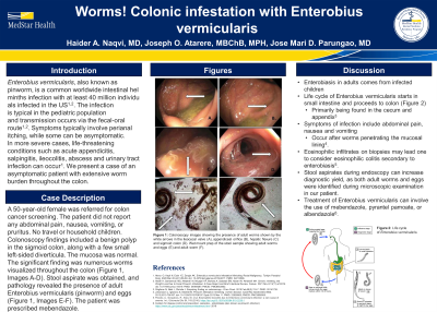
Case Description/Methods: A 50-year-old female was referred for colon cancer screening. The patient did not report any abdominal pain, nausea, vomiting, or pruritus. No travel or household children. Colonoscopy findings included a benign polyp in the sigmoid colon, along with a few small left-sided diverticula. The mucosa was normal. The significant finding was numerous worms visualized throughout the colon (Figure 1). Stool aspirate was obtained, and pathology revealed the presence of adult Enterobius vermicularis (pinworm) and eggs (Figure 2). The patient was prescribed mebendazole.
Discussion: Adults with enterobiasis tend to get infected from children in the household, following transmission from other children. The life cycle of Enterobius vermicularis leads to the eggs releasing their larvae into the small intestines, and adult worms migrate to the colon, primarily the cecum and appendix3. Adult worms in our patient were found in the cecum and throughout the colon, with extensive disease burden. Such a high disease burden can lead to abdominal pain, nausea and vomiting; our patient was asymptomatic. GI symptoms occur in patients with worms penetrating the mucosal lining secondary to any additional insult4. The infiltration of the mucosal lining leads to possible life-threatening conditions. Eosinophilic infiltrates on biopsies may lead one to consider eosinophilic colitis secondary to enterobiasis5. Despite the high worm burden, the absence of mucosal damage led to our patient having no symptoms. Diagnosis is challenging, as eggs are usually found in the perineum, which can be missed by a typical stool collection. Stool aspirates during endoscopy can increase diagnostic yield, as both adult worms and eggs were identified during microscopic examination in our patient. Treatment of Enterobius vermicularis can involve the use of mebendazole, pyrantel pamoate, or albendazole6.

Disclosures:
Haider A. Naqvi, MD1, Joseph O. Atarere, MBChB, MPH2, Jose Mari D. Parungao, MD3. P3088 - Worms! Colonic Infestation with Enterobius Vermicularis, ACG 2023 Annual Scientific Meeting Abstracts. Vancouver, BC, Canada: American College of Gastroenterology.
1MedStar Health, Baltimore, MD; 2MedStar Union Memorial Hospital, Baltimore, MD; 3MedStar Franklin Square Medical Center, Baltimore, MD
Introduction: Enterobius vermicularis, also known as pinworm, is a common worldwide intestinal helminths infection with at least 40 million individuals infected in the US1,2. The infection is typical in the pediatric population and transmission occurs via the fecal-oral route1,2. Symptoms typically involve perianal itching, while some can be asymptomatic. In more severe cases, life-threatening conditions such as acute appendicitis, salpingitis, ileocolitis, abscess and urinary tract infection can occur1. We present a case of an asymptomatic patient with extensive worm burden throughout the colon.
Case Description/Methods: A 50-year-old female was referred for colon cancer screening. The patient did not report any abdominal pain, nausea, vomiting, or pruritus. No travel or household children. Colonoscopy findings included a benign polyp in the sigmoid colon, along with a few small left-sided diverticula. The mucosa was normal. The significant finding was numerous worms visualized throughout the colon (Figure 1). Stool aspirate was obtained, and pathology revealed the presence of adult Enterobius vermicularis (pinworm) and eggs (Figure 2). The patient was prescribed mebendazole.
Discussion: Adults with enterobiasis tend to get infected from children in the household, following transmission from other children. The life cycle of Enterobius vermicularis leads to the eggs releasing their larvae into the small intestines, and adult worms migrate to the colon, primarily the cecum and appendix3. Adult worms in our patient were found in the cecum and throughout the colon, with extensive disease burden. Such a high disease burden can lead to abdominal pain, nausea and vomiting; our patient was asymptomatic. GI symptoms occur in patients with worms penetrating the mucosal lining secondary to any additional insult4. The infiltration of the mucosal lining leads to possible life-threatening conditions. Eosinophilic infiltrates on biopsies may lead one to consider eosinophilic colitis secondary to enterobiasis5. Despite the high worm burden, the absence of mucosal damage led to our patient having no symptoms. Diagnosis is challenging, as eggs are usually found in the perineum, which can be missed by a typical stool collection. Stool aspirates during endoscopy can increase diagnostic yield, as both adult worms and eggs were identified during microscopic examination in our patient. Treatment of Enterobius vermicularis can involve the use of mebendazole, pyrantel pamoate, or albendazole6.

Figure: Image 1: Colonoscopy images showing the presence of adult worms shown by the white arrows in the ileocecal valve (A), appendiceal orifice (B), hepatic flexure (C) and sigmoid colon (D). Wet mount prep of the stool sample showing adult worms and eggs (E) and adult worm (F).
Disclosures:
Haider Naqvi indicated no relevant financial relationships.
Joseph Atarere indicated no relevant financial relationships.
Jose Mari Parungao indicated no relevant financial relationships.
Haider A. Naqvi, MD1, Joseph O. Atarere, MBChB, MPH2, Jose Mari D. Parungao, MD3. P3088 - Worms! Colonic Infestation with Enterobius Vermicularis, ACG 2023 Annual Scientific Meeting Abstracts. Vancouver, BC, Canada: American College of Gastroenterology.
