Tuesday Poster Session
Category: Colon
P3093 - Insights Through the Endoscope: Unveiling the Endoscopic Features of Colorectal Follicular Lymphoma
Tuesday, October 24, 2023
10:30 AM - 4:00 PM PT
Location: Exhibit Hall

Has Audio
- JG
Jacob J. Gries, MD, MSc
Geisinger Medical Center
Danville, PA
Presenting Author(s)
Jacob J.. Gries, MD, MSc1, Bing Chen, MD1, Duane E. Deivert, DO, FACG2, Liping Li, MD, PhD1
1Geisinger Medical Center, Danville, PA; 2Geisinger Northeast, Wilkes-Barre, PA
Introduction: Primary colorectal lymphoma (PCL) is a very rare disease, accounting for 0.2-0.6% of all colorectal malignancies. The disease is typically identified on routine colonoscopy and has an indolent course1. Its rarity limits the available information about its macroscopic features; however, existing studies have suggested a wide variety of types (polypoid, ulcerative, multiple lymphomatous polyposis, diffuse, and mixed). The distribution of these lesions also varies with the majority being located in the cecum (30-60%) followed by the sigmoid colon and rectum (10-25%)2,3. This case of early-stage PCL aims to contribute to the limited existing literature regarding the endoscopic features and distribution of PCL.
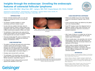
Case Description/Methods: The patient is a 69-year-old female with remote history of tobacco abuse and remote history of villous adenoma who underwent surveillance colonoscopy revealing one 8 mm sessile polyp in the ascending colon, five 3-6 mm polyps in the transverse colon, and two 4 mm sessile polyps in the rectum (Figure 1). All polyps were removed with a cold snare. The pathologies of the ascending colon and rectal polyps were consistent with grade 1-2/3 and grade 1/3 follicular lymphoma, respectively (Figure 2). Two Deauville 5 lymph nodes (aortocaval and left inguinal) were identified on PET scan. Patient was stratified as low risk via the Follicular Lymphoma International Prognostic Index (FLIPI) score. A trial of rituximab therapy was deferred by the patient due to risk of adverse effects. Serial PET imaging 2 years from diagnosis found no significant change in lymph node involvement, and follow-up colonoscopy observed no recurrence or persistent lymphoma.
Discussion: After 2 years of follow-up, our patient has not had any evidence of progression consistent with the typical indolent nature of this disease. With one sessile polyp in the ascending colon and two sessile polyps in the rectum, this data concurs with current literature about the macroscopic features and distribution of PCL lesions. Further studies to establish distinctive features and distribution of PCL lesions in conjunction with histopathological evaluation may enhance diagnosis and aid in effective management of this unique colorectal neoplasm.

Disclosures:
Jacob J.. Gries, MD, MSc1, Bing Chen, MD1, Duane E. Deivert, DO, FACG2, Liping Li, MD, PhD1. P3093 - Insights Through the Endoscope: Unveiling the Endoscopic Features of Colorectal Follicular Lymphoma, ACG 2023 Annual Scientific Meeting Abstracts. Vancouver, BC, Canada: American College of Gastroenterology.
1Geisinger Medical Center, Danville, PA; 2Geisinger Northeast, Wilkes-Barre, PA
Introduction: Primary colorectal lymphoma (PCL) is a very rare disease, accounting for 0.2-0.6% of all colorectal malignancies. The disease is typically identified on routine colonoscopy and has an indolent course1. Its rarity limits the available information about its macroscopic features; however, existing studies have suggested a wide variety of types (polypoid, ulcerative, multiple lymphomatous polyposis, diffuse, and mixed). The distribution of these lesions also varies with the majority being located in the cecum (30-60%) followed by the sigmoid colon and rectum (10-25%)2,3. This case of early-stage PCL aims to contribute to the limited existing literature regarding the endoscopic features and distribution of PCL.
Case Description/Methods: The patient is a 69-year-old female with remote history of tobacco abuse and remote history of villous adenoma who underwent surveillance colonoscopy revealing one 8 mm sessile polyp in the ascending colon, five 3-6 mm polyps in the transverse colon, and two 4 mm sessile polyps in the rectum (Figure 1). All polyps were removed with a cold snare. The pathologies of the ascending colon and rectal polyps were consistent with grade 1-2/3 and grade 1/3 follicular lymphoma, respectively (Figure 2). Two Deauville 5 lymph nodes (aortocaval and left inguinal) were identified on PET scan. Patient was stratified as low risk via the Follicular Lymphoma International Prognostic Index (FLIPI) score. A trial of rituximab therapy was deferred by the patient due to risk of adverse effects. Serial PET imaging 2 years from diagnosis found no significant change in lymph node involvement, and follow-up colonoscopy observed no recurrence or persistent lymphoma.
Discussion: After 2 years of follow-up, our patient has not had any evidence of progression consistent with the typical indolent nature of this disease. With one sessile polyp in the ascending colon and two sessile polyps in the rectum, this data concurs with current literature about the macroscopic features and distribution of PCL lesions. Further studies to establish distinctive features and distribution of PCL lesions in conjunction with histopathological evaluation may enhance diagnosis and aid in effective management of this unique colorectal neoplasm.

Figure: A&B) One medium-sized sessile polypoid lesion located in the ascending colon and two small sessile polypoid lesions located in the rectum. C&D) H&E staining showed back to back secondary follicles with geminal center at 20x and 100x. E) IHC staining showed BCL2 positive in geminal centers at 20x.
Disclosures:
Jacob Gries indicated no relevant financial relationships.
Bing Chen indicated no relevant financial relationships.
Duane Deivert indicated no relevant financial relationships.
Liping Li indicated no relevant financial relationships.
Jacob J.. Gries, MD, MSc1, Bing Chen, MD1, Duane E. Deivert, DO, FACG2, Liping Li, MD, PhD1. P3093 - Insights Through the Endoscope: Unveiling the Endoscopic Features of Colorectal Follicular Lymphoma, ACG 2023 Annual Scientific Meeting Abstracts. Vancouver, BC, Canada: American College of Gastroenterology.
