Tuesday Poster Session
Category: Colon
P3099 - Intussusception From a Giant Colon Lipoma
Tuesday, October 24, 2023
10:30 AM - 4:00 PM PT
Location: Exhibit Hall

Has Audio

Naveena Sunkara, MD
Warren Alpert Medical School, Brown University
Providence, RI
Presenting Author(s)
Naveena Sunkara, MD1, Lindsey Channen, MD2, Colleen R. Kelly, MD, FACG3
1Warren Alpert Medical School, Brown University, Providence, RI; 2Warren Alpert Medical School of Brown University, Providence, RI; 3Alpert School of Medicine of Brown University, Providence, RI
Introduction: Colon lipomas are benign submucosal lesions which are asymptomatic in a majority of cases. Though rare, complications from lipomas can be serious including hemorrhage and intestinal obstruction. Here we present a case of intussusception caused by a giant colon lipoma.
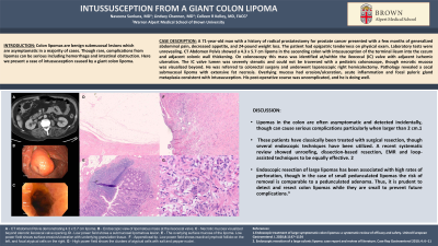
Case Description/Methods: A 71-year-old man with a history of radical prostatectomy for prostate cancer presented with a few months of generalized abdominal pain, decreased appetite, and 24-pound weight loss. The patient had epigastric tenderness on physical exam. Laboratory tests were unrevealing. CT Abdomen Pelvis showed a 4.3 x 5.7 cm lipoma in the ascending colon with intussusception of the terminal ileum into the cecum and adjacent colonic wall thickening. On colonoscopy this mass was identified at/within the ileocecal (IC) valve with adjacent ischemic ulceration. The IC valve lumen was severely stenotic and could not be traversed with a pediatric colonoscope, though necrotic mucosa was visualized beyond. He was referred to colorectal surgery and underwent laparoscopic right hemicolectomy. Pathology revealed a cecal submucosal lipoma with extensive fat necrosis. Overlying mucosa had erosion/ulceration, acute inflammation and focal pyloric gland metaplasia consistent with intussusception. His post-operative course was uncomplicated, and he is doing well.
Discussion: Lipomas in the colon are often asymptomatic and detected incidentally, though can cause serious complications particularly when larger than 2 cm. These patients have classically been treated with surgical resection, though several endoscopic techniques have been utilized. A recent systematic review showed unroofing, dissection-based resection, EMR and loop-assisted techniques to be equally effective. Endoscopic resection of large lipomas has been associated with high rates of perforation, though in the case of small pedunculated lipomas the risk of removal is comparable to a pedunculated adenoma. Thus, it is prudent to detect and resect colon lipomas while they are small to prevent future complications.

Disclosures:
Naveena Sunkara, MD1, Lindsey Channen, MD2, Colleen R. Kelly, MD, FACG3. P3099 - Intussusception From a Giant Colon Lipoma, ACG 2023 Annual Scientific Meeting Abstracts. Vancouver, BC, Canada: American College of Gastroenterology.
1Warren Alpert Medical School, Brown University, Providence, RI; 2Warren Alpert Medical School of Brown University, Providence, RI; 3Alpert School of Medicine of Brown University, Providence, RI
Introduction: Colon lipomas are benign submucosal lesions which are asymptomatic in a majority of cases. Though rare, complications from lipomas can be serious including hemorrhage and intestinal obstruction. Here we present a case of intussusception caused by a giant colon lipoma.
Case Description/Methods: A 71-year-old man with a history of radical prostatectomy for prostate cancer presented with a few months of generalized abdominal pain, decreased appetite, and 24-pound weight loss. The patient had epigastric tenderness on physical exam. Laboratory tests were unrevealing. CT Abdomen Pelvis showed a 4.3 x 5.7 cm lipoma in the ascending colon with intussusception of the terminal ileum into the cecum and adjacent colonic wall thickening. On colonoscopy this mass was identified at/within the ileocecal (IC) valve with adjacent ischemic ulceration. The IC valve lumen was severely stenotic and could not be traversed with a pediatric colonoscope, though necrotic mucosa was visualized beyond. He was referred to colorectal surgery and underwent laparoscopic right hemicolectomy. Pathology revealed a cecal submucosal lipoma with extensive fat necrosis. Overlying mucosa had erosion/ulceration, acute inflammation and focal pyloric gland metaplasia consistent with intussusception. His post-operative course was uncomplicated, and he is doing well.
Discussion: Lipomas in the colon are often asymptomatic and detected incidentally, though can cause serious complications particularly when larger than 2 cm. These patients have classically been treated with surgical resection, though several endoscopic techniques have been utilized. A recent systematic review showed unroofing, dissection-based resection, EMR and loop-assisted techniques to be equally effective. Endoscopic resection of large lipomas has been associated with high rates of perforation, though in the case of small pedunculated lipomas the risk of removal is comparable to a pedunculated adenoma. Thus, it is prudent to detect and resect colon lipomas while they are small to prevent future complications.

Figure: A - CT Abdomen Pelvis demonstrating 4.3 x 5.7 cm lipoma.
B - Endoscopic view of lipomatous mass at the ileocecal valve.
C - Necrotic mucosa visualized beyond stenotic ileocecal valve opening.
D - Low power field shows a submucosal lipomatous lesion.
E - The overlying surface mucosa of the lipoma. Low power field shows surface erosion/ulceration with underlying granulation tissue.
F - Appendiceal tip. Low power field shows reactive lymphoid follicle on the left, and focal atypical cells on the right.
G - High power field shows the clusters of atypical cells with salt and pepper nuclei .
Disclosures:
Naveena Sunkara indicated no relevant financial relationships.
Lindsey Channen indicated no relevant financial relationships.
Colleen Kelly: Sebela Pharmaceuticals – Consultant.
Naveena Sunkara, MD1, Lindsey Channen, MD2, Colleen R. Kelly, MD, FACG3. P3099 - Intussusception From a Giant Colon Lipoma, ACG 2023 Annual Scientific Meeting Abstracts. Vancouver, BC, Canada: American College of Gastroenterology.
