Tuesday Poster Session
Category: Colon
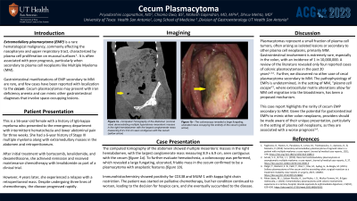
P3111 - Cecum Plasmacytoma
Tuesday, October 24, 2023
10:30 AM - 4:00 PM PT
Location: Exhibit Hall

Has Audio

Priyadarshini Loganathan, MD
University of Texas Health Science Center
San Antonio, TX
Presenting Author(s)
Priyadarshini Loganathan, MD1, Chioma Owo, BS2, Mahesh Gajendran, MD, MPH3, Dhruv Mehta, MD2
1University of Texas Health Science Center, San Antonio, TX; 2UT Health San Antonio, San Antonio, TX; 3University of Texas Health Science Center at San Antonio, San Antonio, TX
Introduction: Extramedullary plasmacytoma (EMP) is a rare hematological malignancy characterized by plasma cell proliferation on mucosal surfaces and is often associated with poor prognosis, particularly when secondary to plasma cell neoplasms like Multiple Myeloma (MM).
Case Description/Methods: This is a 56-year-old female with a history of IgG-kappa myeloma presented to the emergency department with intermittent hematochezia and lower abdominal pain for three weeks. She had a 6-year history of Stage III multiple myeloma along with extramedullary masses in the abdomen and retroperitoneum. After initial treatment with Bortezomib, lenalidomide, and dexamethasone, she achieved remission and received maintenance chemotherapy with lenalidomide as part of a clinical trial. However, 4 years later, she experienced a relapse with a retroperitoneal mass. Despite undergoing three lines of chemotherapy, the disease progressed rapidly. The computed tomography (CT) of the abdomen showed multiple mesenteric masses in the right hemiabdomen, with the largest conglomerate mass measuring 8.9 x 6.9 cm, seen contiguous with the cecum. To further evaluate hematochezia, a colonoscopy was performed, which revealed a large fungating, ulcerated, friable mass in the cecum confirmed to be plasmacytoma with anaplastic features. Immunohistochemistry showed positivity for CD138 and MUM-1 with kappa light chain restriction. The patient was started on palliative chemotherapy, but her condition continued to worsen, leading to the decision for hospice care, and she eventually succumbed to the disease.
Discussion: Plasmacytomas represent a small fraction of plasma cell tumors, often arising as isolated lesions or secondary to other plasma cell neoplasms, primarily multiple myeloma. Gastrointestinal involvement is extremely rare, especially in the colon, with an incidence of 1 in 10,000,000. A review of the literature revealed only four reported cases of colonic plasmacytomas in the past 20 years. The pathophysiology of EMPs is undetermined, in the setting of MM, “plasma cell escape”, where extracellular matrix alterations allow for MM cell migration into the bloodstream, has been a proposed mechanism. This case report highlights the rarity of cecum EMP secondary to MM. Given the potential for gastrointestinal EMPs to mimic other colon neoplasms, providers should be made aware of their unique presentation, particularly in the setting of plasma cell neoplasms, as they are associated with a worse prognosis.

Disclosures:
Priyadarshini Loganathan, MD1, Chioma Owo, BS2, Mahesh Gajendran, MD, MPH3, Dhruv Mehta, MD2. P3111 - Cecum Plasmacytoma, ACG 2023 Annual Scientific Meeting Abstracts. Vancouver, BC, Canada: American College of Gastroenterology.
1University of Texas Health Science Center, San Antonio, TX; 2UT Health San Antonio, San Antonio, TX; 3University of Texas Health Science Center at San Antonio, San Antonio, TX
Introduction: Extramedullary plasmacytoma (EMP) is a rare hematological malignancy characterized by plasma cell proliferation on mucosal surfaces and is often associated with poor prognosis, particularly when secondary to plasma cell neoplasms like Multiple Myeloma (MM).
Case Description/Methods: This is a 56-year-old female with a history of IgG-kappa myeloma presented to the emergency department with intermittent hematochezia and lower abdominal pain for three weeks. She had a 6-year history of Stage III multiple myeloma along with extramedullary masses in the abdomen and retroperitoneum. After initial treatment with Bortezomib, lenalidomide, and dexamethasone, she achieved remission and received maintenance chemotherapy with lenalidomide as part of a clinical trial. However, 4 years later, she experienced a relapse with a retroperitoneal mass. Despite undergoing three lines of chemotherapy, the disease progressed rapidly. The computed tomography (CT) of the abdomen showed multiple mesenteric masses in the right hemiabdomen, with the largest conglomerate mass measuring 8.9 x 6.9 cm, seen contiguous with the cecum. To further evaluate hematochezia, a colonoscopy was performed, which revealed a large fungating, ulcerated, friable mass in the cecum confirmed to be plasmacytoma with anaplastic features. Immunohistochemistry showed positivity for CD138 and MUM-1 with kappa light chain restriction. The patient was started on palliative chemotherapy, but her condition continued to worsen, leading to the decision for hospice care, and she eventually succumbed to the disease.
Discussion: Plasmacytomas represent a small fraction of plasma cell tumors, often arising as isolated lesions or secondary to other plasma cell neoplasms, primarily multiple myeloma. Gastrointestinal involvement is extremely rare, especially in the colon, with an incidence of 1 in 10,000,000. A review of the literature revealed only four reported cases of colonic plasmacytomas in the past 20 years. The pathophysiology of EMPs is undetermined, in the setting of MM, “plasma cell escape”, where extracellular matrix alterations allow for MM cell migration into the bloodstream, has been a proposed mechanism. This case report highlights the rarity of cecum EMP secondary to MM. Given the potential for gastrointestinal EMPs to mimic other colon neoplasms, providers should be made aware of their unique presentation, particularly in the setting of plasma cell neoplasms, as they are associated with a worse prognosis.

Figure: Figure 1a- Computed Tomography of the Abdomen (coronal view) demonstrating multiple hyperdense mesenteric masses in the right hemiabdomen with the largest conglomerate mass measuring 8.9 x 6.9 cm seen contiguous with the cecum (yellow arrow)
Figure 1b- The colonoscopy revealed a large fungating, ulcerated mass occupying the entirety of the cecum (yellow arrow).
Figure 1b- The colonoscopy revealed a large fungating, ulcerated mass occupying the entirety of the cecum (yellow arrow).
Disclosures:
Priyadarshini Loganathan indicated no relevant financial relationships.
Chioma Owo indicated no relevant financial relationships.
Mahesh Gajendran indicated no relevant financial relationships.
Dhruv Mehta indicated no relevant financial relationships.
Priyadarshini Loganathan, MD1, Chioma Owo, BS2, Mahesh Gajendran, MD, MPH3, Dhruv Mehta, MD2. P3111 - Cecum Plasmacytoma, ACG 2023 Annual Scientific Meeting Abstracts. Vancouver, BC, Canada: American College of Gastroenterology.
