Tuesday Poster Session
Category: Colon
P3112 - A Rare Case of Primary Gastrointestinal Burkitt’s Lymphoma With Rapid Progression
Tuesday, October 24, 2023
10:30 AM - 4:00 PM PT
Location: Exhibit Hall

Has Audio

Priya Krishnaprasad, DO
Christiana Care Health System
Newark, DE
Presenting Author(s)
Priya Krishnaprasad, DO, Mai Lam, DO
Christiana Care Health System, Newark, DE
Introduction: Primary gastrointestinal non-Hodgkin’s lymphomas, such as Burkitt’s lymphoma, are extremely rare accounting for only about 0.9% of all gastrointestinal tumors and less than 0.5% of colonic malignancies. This case highlights the tendency of Burkitt’s lymphoma to rapidly progress. Additionally, it highlights key features that are commonly seen in patients that have this rare tumor including prior history of chemotherapy or radiotherapy and EBV positivity. It is important for clinicians to recognize this disease in order to rapidly initiate treatment for these patients given its poor prognosis.
Case Description/Methods: This case pertains to a 38-year-old Egyptian male with a history of Hodgkin's lymphoma treated with chemotherapy in 2015 who presented to the ED with 3 days of diffuse abdominal pain. He reported not having a bowel movement for over 4 days, along with unintentional weight loss of 8 kg within 2 months. Physical exam revealed minimal right lower quadrant tenderness without distention, rigidity or guarding. Labs showed mildly elevated liver function tests, and negative lipase. Decision was made to obtain imaging which revealed a solid mass in the ileocecal junction, raising concern for neoplasm. Additionally, multiple hepatic lesions, suggesting metastatic disease was also observed. No masses or lesions were seen on CT abdomen pelvis imaging from just one month ago suggesting extremely rapid progression. Colonoscopy confirmed a non-obstructing polypoid and submucosal mass at the ileocecal valve which measured 5 cm in length. Biopsies from colonoscopy and image guided liver biopsy were completed and pathology results favored high-grade Burkitt's lymphoma. He was seen to also be CMV IgG and EBV IgG positive. The patient underwent further diagnostics for staging and was started on chemotherapy per oncology.
Discussion: Primary gastrointestinal Burkitt’s lymphoma is rare and predominantly shows up in the ileocecal junction. Tumor growth tends to be rapid as seen in this patient who developed a tumor along with hepatic metastasis within one month. Clinicians should maintain a low threshold of suspicion for Burkitt’s lymphoma in patients who have previously undergone chemotherapy and present with a mass in the ileocecal junction. Additionally, a history of EBV infection, as seen in this case, can be a predisposing factor. Early detection and treatment are crucial to mitigate the exceedingly high mortality risk that unfortunately exists for these patients.

Disclosures:
Priya Krishnaprasad, DO, Mai Lam, DO. P3112 - A Rare Case of Primary Gastrointestinal Burkitt’s Lymphoma With Rapid Progression, ACG 2023 Annual Scientific Meeting Abstracts. Vancouver, BC, Canada: American College of Gastroenterology.
Christiana Care Health System, Newark, DE
Introduction: Primary gastrointestinal non-Hodgkin’s lymphomas, such as Burkitt’s lymphoma, are extremely rare accounting for only about 0.9% of all gastrointestinal tumors and less than 0.5% of colonic malignancies. This case highlights the tendency of Burkitt’s lymphoma to rapidly progress. Additionally, it highlights key features that are commonly seen in patients that have this rare tumor including prior history of chemotherapy or radiotherapy and EBV positivity. It is important for clinicians to recognize this disease in order to rapidly initiate treatment for these patients given its poor prognosis.
Case Description/Methods: This case pertains to a 38-year-old Egyptian male with a history of Hodgkin's lymphoma treated with chemotherapy in 2015 who presented to the ED with 3 days of diffuse abdominal pain. He reported not having a bowel movement for over 4 days, along with unintentional weight loss of 8 kg within 2 months. Physical exam revealed minimal right lower quadrant tenderness without distention, rigidity or guarding. Labs showed mildly elevated liver function tests, and negative lipase. Decision was made to obtain imaging which revealed a solid mass in the ileocecal junction, raising concern for neoplasm. Additionally, multiple hepatic lesions, suggesting metastatic disease was also observed. No masses or lesions were seen on CT abdomen pelvis imaging from just one month ago suggesting extremely rapid progression. Colonoscopy confirmed a non-obstructing polypoid and submucosal mass at the ileocecal valve which measured 5 cm in length. Biopsies from colonoscopy and image guided liver biopsy were completed and pathology results favored high-grade Burkitt's lymphoma. He was seen to also be CMV IgG and EBV IgG positive. The patient underwent further diagnostics for staging and was started on chemotherapy per oncology.
Discussion: Primary gastrointestinal Burkitt’s lymphoma is rare and predominantly shows up in the ileocecal junction. Tumor growth tends to be rapid as seen in this patient who developed a tumor along with hepatic metastasis within one month. Clinicians should maintain a low threshold of suspicion for Burkitt’s lymphoma in patients who have previously undergone chemotherapy and present with a mass in the ileocecal junction. Additionally, a history of EBV infection, as seen in this case, can be a predisposing factor. Early detection and treatment are crucial to mitigate the exceedingly high mortality risk that unfortunately exists for these patients.

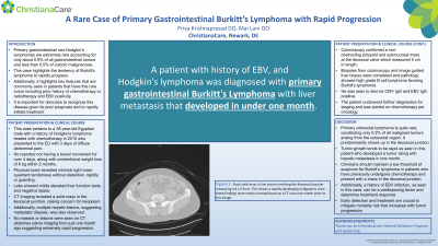
Figure: This CT abdomen pelvis image reveals a focal solid mass at the cecum involving the ileocecal junction which was later confirmed to be Burkitt's Lymphoma.
Disclosures:
Priya Krishnaprasad indicated no relevant financial relationships.
Mai Lam indicated no relevant financial relationships.
Priya Krishnaprasad, DO, Mai Lam, DO. P3112 - A Rare Case of Primary Gastrointestinal Burkitt’s Lymphoma With Rapid Progression, ACG 2023 Annual Scientific Meeting Abstracts. Vancouver, BC, Canada: American College of Gastroenterology.
