Tuesday Poster Session
Category: Small Intestine
P4127 - An Unusual Case of Small Bowel Obstruction in a Virgin Abdomen
Tuesday, October 24, 2023
10:30 AM - 4:00 PM PT
Location: Exhibit Hall

Has Audio
- IB
Ifrah Z. Butt, MD
HCA Florida Aventura Hospital
Aventura, FL
Presenting Author(s)
Ifrah Z. Butt, MD, Mina Ayad, MD, Ramasamy S. Nathan, MD
HCA Florida Aventura Hospital, Aventura, FL
Introduction: Small bowel neuroendocrine tumors (SBNETs) arise from the enterochromaffin cells that line the gastrointestinal tract and secrete serotonin. Although considered rare, SBNETs have been increasing in incidence and prevalence, likely due to the increase in imaging studies. We present a case of an elderly man who presented with a small bowel obstruction and was found to have a multicentric SBNET in the mid-ileum.
Case Description/Methods: An 85-year-old male presented to the emergency department with complaints of diffuse abdominal pain, nausea, and vomiting for the past 2 days. He had no relevant medical conditions, no history of prior abdominal surgeries and was up to date on his colonoscopy. On admission, he was tachycardic and hypertensive. His physical exam revealed a distended and tympanic abdomen with diffuse tenderness to palpation and hypoactive bowel sounds. His WBC was 16,300 cells/uL. A computed tomography (CT) scan of the abdomen and pelvis revealed diffusely dilated small bowel loops with air-fluid levels and a transition point in the right lower quadrant. In the region of the transition point, there was a nodular mass-like structure measuring 1.8 x 1.5 cm, in addition to slight nodularity and stranding of several areas of the omentum. Since conservative nasogastric tube (NGT) decompression failed, he was taken for exploratory laparotomy. A mid-small bowel mass with obstruction was discovered, with a tight band tethering the mesentery. Surgical resection with primary anastomosis as well as lymph node and mesentery dissection were performed. Pathology report showed two foci of well-differentiated small bowel neuroendocrine tumors measuring 1.6 and 2 cm in the greatest dimension, Grade 2, with evidence of invasion through muscularis propria and subserosal tissue. There was also lymphatic and perineural invasion with one out of two lymph nodes involved. Pankeratin, chromogranin and synaptophysin tumor stains were positive. The Ki-67 index was 5-10%. Chromogranin A level was elevated. The patient did well post-surgery and was referred for outpatient oncological follow-up.
Discussion: Small bowel tumors are rare and correspond to 1-2% of all gastrointestinal malignancies and SBNETs comprise of only a subset of these rare neoplasms. Usually SBNETs are small and multicentric located in the distal ileum. However, despite the small size, there is still a high risk of lymph node and distal metastasis, similar to our case.

Disclosures:
Ifrah Z. Butt, MD, Mina Ayad, MD, Ramasamy S. Nathan, MD. P4127 - An Unusual Case of Small Bowel Obstruction in a Virgin Abdomen, ACG 2023 Annual Scientific Meeting Abstracts. Vancouver, BC, Canada: American College of Gastroenterology.
HCA Florida Aventura Hospital, Aventura, FL
Introduction: Small bowel neuroendocrine tumors (SBNETs) arise from the enterochromaffin cells that line the gastrointestinal tract and secrete serotonin. Although considered rare, SBNETs have been increasing in incidence and prevalence, likely due to the increase in imaging studies. We present a case of an elderly man who presented with a small bowel obstruction and was found to have a multicentric SBNET in the mid-ileum.
Case Description/Methods: An 85-year-old male presented to the emergency department with complaints of diffuse abdominal pain, nausea, and vomiting for the past 2 days. He had no relevant medical conditions, no history of prior abdominal surgeries and was up to date on his colonoscopy. On admission, he was tachycardic and hypertensive. His physical exam revealed a distended and tympanic abdomen with diffuse tenderness to palpation and hypoactive bowel sounds. His WBC was 16,300 cells/uL. A computed tomography (CT) scan of the abdomen and pelvis revealed diffusely dilated small bowel loops with air-fluid levels and a transition point in the right lower quadrant. In the region of the transition point, there was a nodular mass-like structure measuring 1.8 x 1.5 cm, in addition to slight nodularity and stranding of several areas of the omentum. Since conservative nasogastric tube (NGT) decompression failed, he was taken for exploratory laparotomy. A mid-small bowel mass with obstruction was discovered, with a tight band tethering the mesentery. Surgical resection with primary anastomosis as well as lymph node and mesentery dissection were performed. Pathology report showed two foci of well-differentiated small bowel neuroendocrine tumors measuring 1.6 and 2 cm in the greatest dimension, Grade 2, with evidence of invasion through muscularis propria and subserosal tissue. There was also lymphatic and perineural invasion with one out of two lymph nodes involved. Pankeratin, chromogranin and synaptophysin tumor stains were positive. The Ki-67 index was 5-10%. Chromogranin A level was elevated. The patient did well post-surgery and was referred for outpatient oncological follow-up.
Discussion: Small bowel tumors are rare and correspond to 1-2% of all gastrointestinal malignancies and SBNETs comprise of only a subset of these rare neoplasms. Usually SBNETs are small and multicentric located in the distal ileum. However, despite the small size, there is still a high risk of lymph node and distal metastasis, similar to our case.

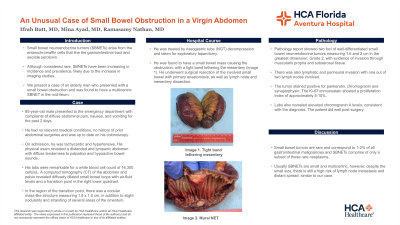
Figure: Resected portion of small bowel with a tight band compressing on the right side
Disclosures:
Ifrah Butt indicated no relevant financial relationships.
Mina Ayad indicated no relevant financial relationships.
Ramasamy Nathan indicated no relevant financial relationships.
Ifrah Z. Butt, MD, Mina Ayad, MD, Ramasamy S. Nathan, MD. P4127 - An Unusual Case of Small Bowel Obstruction in a Virgin Abdomen, ACG 2023 Annual Scientific Meeting Abstracts. Vancouver, BC, Canada: American College of Gastroenterology.
