Tuesday Poster Session
Category: Colon
P3145 - A Rare Occurrence of Basaloid Squamous Cell Carcinoma of Rectum
Tuesday, October 24, 2023
10:30 AM - 4:00 PM PT
Location: Exhibit Hall

Has Audio

Harshavardhan Sanekommu, MD
Hackensack Meridian Jersey Shore University Medical Center
Neptune, NJ
Presenting Author(s)
Harshavardhan Sanekommu, MD1, Mohammed Abdila, MD2, Karthikeyan Swaminath, MD3, Sobaan Taj, MD1, Jayasree Ravilla, MD4, Mohammad Hossain, MD2
1Hackensack Meridian Jersey Shore University Medical Center, Neptune, NJ; 2Hackensack Meridian Jersey Shore University Medical Center, Neptune Township, NJ; 3Hackensack Meridian JFK Medical Center, Edison, NJ; 4Monmouth Medical Center/RWJBH, Long Branch, NJ
Introduction: Squamous cell carcinomas (SCC) are the prevailing form of tumors found in the anal canal and perianal region, with the basaloid variant being the most frequently observed. Basaloid SCC of the rectum is extremely rare and only reported 10 times. Here, we report this rare case of basaloid SCC of the rectum and the associated poor prognosis.
Case Description/Methods: 66-year-old with no significant past medical history presented to the emergency department with fatigue and intermittent hematochezia for the last eight months. She denied weight loss, night sweats/chills, loss of appetite, and use of nonsteroidal anti-inflammatory drugs. Patient hasn’t followed up with a primary care physician for many years. She is not up to date with her screening, including screening for colon cancer. In the emergency department, her vitals were stable. Physical exam revealed pale skin and conjunctiva, with a benign abdomen. Direct rectal exam showed light brown stool in the rectal vault with a palpable nodular mass and a normal rectal tone. Her labs were significant for: hemoglobin 3.9, hematocrit 16.0, and mean corpuscular volume 71.1. Fecal occult blood test was positive. Patient was given two units of packed red blood cells and post-transfusion hemoglobin was 8.5. The following day, the patient underwent Esophagogastroduodenoscopy (EGD) and colonoscopy. EGD showed a Z-line that was slightly irregular and the of the stomach and duodenum was unremarkable. The colonoscopy revealed a fungating non-obstructing large mass in the distal rectum (Image 1), and scattered diverticula in the sigmoid and descending colon without evidence of recent bleeding. The rectal mass was biopsied, which later showed poorly differentiated carcinoma with basaloid features, supportive of basaloid squamous cell carcinoma of rectum. Magnetic Resonance Imaging of the pelvis revealed circumferential thickening within the rectum and tumor extension beyond the muscularis propria, into the mesorectal fat suggesting cancer staging of T3. The patient is currently undergoing neoadjuvant chemotherapy and radiation therapy with poor response.
Discussion: Basal SCC exhibit a rapid decline due to their heightened aggressiveness compared to other variants of SCC. These Basaloid SCCs are frequently detected and diagnosed in advanced stages, leading to earlier metastasis. Furthermore, BSCCs found in the colorectal region demonstrate unfavorable prognoses and swift progression.

Disclosures:
Harshavardhan Sanekommu, MD1, Mohammed Abdila, MD2, Karthikeyan Swaminath, MD3, Sobaan Taj, MD1, Jayasree Ravilla, MD4, Mohammad Hossain, MD2. P3145 - A Rare Occurrence of Basaloid Squamous Cell Carcinoma of Rectum, ACG 2023 Annual Scientific Meeting Abstracts. Vancouver, BC, Canada: American College of Gastroenterology.
1Hackensack Meridian Jersey Shore University Medical Center, Neptune, NJ; 2Hackensack Meridian Jersey Shore University Medical Center, Neptune Township, NJ; 3Hackensack Meridian JFK Medical Center, Edison, NJ; 4Monmouth Medical Center/RWJBH, Long Branch, NJ
Introduction: Squamous cell carcinomas (SCC) are the prevailing form of tumors found in the anal canal and perianal region, with the basaloid variant being the most frequently observed. Basaloid SCC of the rectum is extremely rare and only reported 10 times. Here, we report this rare case of basaloid SCC of the rectum and the associated poor prognosis.
Case Description/Methods: 66-year-old with no significant past medical history presented to the emergency department with fatigue and intermittent hematochezia for the last eight months. She denied weight loss, night sweats/chills, loss of appetite, and use of nonsteroidal anti-inflammatory drugs. Patient hasn’t followed up with a primary care physician for many years. She is not up to date with her screening, including screening for colon cancer. In the emergency department, her vitals were stable. Physical exam revealed pale skin and conjunctiva, with a benign abdomen. Direct rectal exam showed light brown stool in the rectal vault with a palpable nodular mass and a normal rectal tone. Her labs were significant for: hemoglobin 3.9, hematocrit 16.0, and mean corpuscular volume 71.1. Fecal occult blood test was positive. Patient was given two units of packed red blood cells and post-transfusion hemoglobin was 8.5. The following day, the patient underwent Esophagogastroduodenoscopy (EGD) and colonoscopy. EGD showed a Z-line that was slightly irregular and the of the stomach and duodenum was unremarkable. The colonoscopy revealed a fungating non-obstructing large mass in the distal rectum (Image 1), and scattered diverticula in the sigmoid and descending colon without evidence of recent bleeding. The rectal mass was biopsied, which later showed poorly differentiated carcinoma with basaloid features, supportive of basaloid squamous cell carcinoma of rectum. Magnetic Resonance Imaging of the pelvis revealed circumferential thickening within the rectum and tumor extension beyond the muscularis propria, into the mesorectal fat suggesting cancer staging of T3. The patient is currently undergoing neoadjuvant chemotherapy and radiation therapy with poor response.
Discussion: Basal SCC exhibit a rapid decline due to their heightened aggressiveness compared to other variants of SCC. These Basaloid SCCs are frequently detected and diagnosed in advanced stages, leading to earlier metastasis. Furthermore, BSCCs found in the colorectal region demonstrate unfavorable prognoses and swift progression.

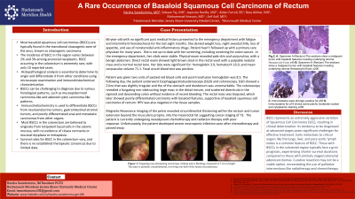
Figure: Figure 1: Fungating non-obstructing rectal mass without active bleeding, measured at 5 cm in length. The mass is partially circumferential, involving one-half of the lumen circumference.
Disclosures:
Harshavardhan Sanekommu indicated no relevant financial relationships.
Mohammed Abdila indicated no relevant financial relationships.
Karthikeyan Swaminath indicated no relevant financial relationships.
Sobaan Taj indicated no relevant financial relationships.
Jayasree Ravilla indicated no relevant financial relationships.
Mohammad Hossain indicated no relevant financial relationships.
Harshavardhan Sanekommu, MD1, Mohammed Abdila, MD2, Karthikeyan Swaminath, MD3, Sobaan Taj, MD1, Jayasree Ravilla, MD4, Mohammad Hossain, MD2. P3145 - A Rare Occurrence of Basaloid Squamous Cell Carcinoma of Rectum, ACG 2023 Annual Scientific Meeting Abstracts. Vancouver, BC, Canada: American College of Gastroenterology.
