Tuesday Poster Session
Category: Small Intestine
P4146 - Metastatic Renal Angiosarcoma to the Small Bowel Causing Recurrent Small Intestinal Bleeds
Tuesday, October 24, 2023
10:30 AM - 4:00 PM PT
Location: Exhibit Hall

Has Audio
- JR
James Rock, DO
Allegheny Health Network
Pittsburgh, PA
Presenting Author(s)
James Rock, DO, Thaer Abdelfattah, MD, MPH, Jeffrey Uchin, MD, Adam Kichler, DO, MEd
Allegheny Health Network, Pittsburgh, PA
Introduction: Angiosarcomas are highly malignant, rare, mesenchymal sarcomas with vascular features. The gold standard for diagnosis is pathological and immunohistochemical confirmation. Angiosarcomas in the small bowel are difficult to diagnose due to late presentation and nonspecific symptoms, such as vomiting and abdominal pain. Primary renal angiosarcoma is even rarer, compared to angiosarcomas of other sites. According to Sabharwal et al., there are only roughly 40 reported cases in literature. Meta-analysis from Zhang et al., revealed that out of 27 of these patient’s studied, none of them had small bowel involvement.
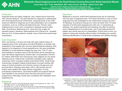
Case Description/Methods: Here, we discuss a 78-year-old male with past medical history of angiosarcoma of the renal pelvis and atrial fibrillation on Xarelto who presented to the hospital with recurrent gastrointestinal bleeding. With regards to his diagnosis of renal angiosarcoma, this was incidentally identified on cross-sectional imaging the year prior. The CT scan demonstrated a complex mass in the left kidney which prompted an open left radical nephrectomy. Months later, he presented to the ED for melena. Both EGD and colonoscopy were negative for any active bleeding source. Patient then underwent outpatient video capsule endoscopy which revealed multiple small bowel AVMs. Small bowel enteroscopy revealed multiple erythematous mucosal nodules with areas of central ulceration and minimal oozing in the jejunum and proximal ileum. Biopsies were taken with a cold forceps and the lesions were treated with argon plasma coagulation. A more protuberant erythematous, semi-pedunculated and polypoid deposit was identified in the proximal ileum that was removed by snare polypectomy. Pathology results were consistent with metastatic renal angiosarcoma.
Discussion: Malignant or primary, small bowel angiosarcomas are an extremely rare sub type of angiosarcomas. This report describes a case of renal angiosarcoma with metastasis to the small bowel causing recurrent GI bleeding. According to Zhang et al, there are fewer than 70 cases reported of small bowel angiosarcoma with only 12 presenting as bleeding. Our patient presented with bleeding only after his primary tumor metastasized to his small bowel, and it was believed that the patient was cancer free prior to presentation. At this point in time, our patient is doing well clinically without further GI bleeding four months following identification of small bowel metastasis and ablative therapy.

Disclosures:
James Rock, DO, Thaer Abdelfattah, MD, MPH, Jeffrey Uchin, MD, Adam Kichler, DO, MEd. P4146 - Metastatic Renal Angiosarcoma to the Small Bowel Causing Recurrent Small Intestinal Bleeds, ACG 2023 Annual Scientific Meeting Abstracts. Vancouver, BC, Canada: American College of Gastroenterology.
Allegheny Health Network, Pittsburgh, PA
Introduction: Angiosarcomas are highly malignant, rare, mesenchymal sarcomas with vascular features. The gold standard for diagnosis is pathological and immunohistochemical confirmation. Angiosarcomas in the small bowel are difficult to diagnose due to late presentation and nonspecific symptoms, such as vomiting and abdominal pain. Primary renal angiosarcoma is even rarer, compared to angiosarcomas of other sites. According to Sabharwal et al., there are only roughly 40 reported cases in literature. Meta-analysis from Zhang et al., revealed that out of 27 of these patient’s studied, none of them had small bowel involvement.
Case Description/Methods: Here, we discuss a 78-year-old male with past medical history of angiosarcoma of the renal pelvis and atrial fibrillation on Xarelto who presented to the hospital with recurrent gastrointestinal bleeding. With regards to his diagnosis of renal angiosarcoma, this was incidentally identified on cross-sectional imaging the year prior. The CT scan demonstrated a complex mass in the left kidney which prompted an open left radical nephrectomy. Months later, he presented to the ED for melena. Both EGD and colonoscopy were negative for any active bleeding source. Patient then underwent outpatient video capsule endoscopy which revealed multiple small bowel AVMs. Small bowel enteroscopy revealed multiple erythematous mucosal nodules with areas of central ulceration and minimal oozing in the jejunum and proximal ileum. Biopsies were taken with a cold forceps and the lesions were treated with argon plasma coagulation. A more protuberant erythematous, semi-pedunculated and polypoid deposit was identified in the proximal ileum that was removed by snare polypectomy. Pathology results were consistent with metastatic renal angiosarcoma.
Discussion: Malignant or primary, small bowel angiosarcomas are an extremely rare sub type of angiosarcomas. This report describes a case of renal angiosarcoma with metastasis to the small bowel causing recurrent GI bleeding. According to Zhang et al, there are fewer than 70 cases reported of small bowel angiosarcoma with only 12 presenting as bleeding. Our patient presented with bleeding only after his primary tumor metastasized to his small bowel, and it was believed that the patient was cancer free prior to presentation. At this point in time, our patient is doing well clinically without further GI bleeding four months following identification of small bowel metastasis and ablative therapy.

Figure: A: Protuberant, erythematous, semi-pedunculated and polypoid deposit
B. Polyp removed via snare polypectomy
C. (200x magnification, H&E) Higher magnification examination shows the lamina propria to contain an infiltrate of atypical cells exhibiting a predominance of plump, fusiform morphology. Rare red blood cells are seen extravasated through the malignant infiltrate.
D. (200x magnification, ERG immunohistochemical stain) Higher magnification of the positive immunoreactivity of the malignant cells for ERG (endothelial marker).
Disclosures:
James Rock indicated no relevant financial relationships.
Thaer Abdelfattah indicated no relevant financial relationships.
Jeffrey Uchin indicated no relevant financial relationships.
Adam Kichler indicated no relevant financial relationships.
James Rock, DO, Thaer Abdelfattah, MD, MPH, Jeffrey Uchin, MD, Adam Kichler, DO, MEd. P4146 - Metastatic Renal Angiosarcoma to the Small Bowel Causing Recurrent Small Intestinal Bleeds, ACG 2023 Annual Scientific Meeting Abstracts. Vancouver, BC, Canada: American College of Gastroenterology.
