Tuesday Poster Session
Category: Colon
P3151 - Inflammatory Cloacogenic Polyp: An Uncommon Cause of Rectal Outlet Bleeding
Tuesday, October 24, 2023
10:30 AM - 4:00 PM PT
Location: Exhibit Hall

Has Audio

Michael A. Perrin, MD, MPH
Baylor College of Medicine
Houston, TX
Presenting Author(s)
Michael A. Perrin, MD, MPH, Linda Green, MD, Rollin George, MD
Baylor College of Medicine, Houston, TX
Introduction: Colonoscopy is often ordered to evaluate rectal outlet bleeding and requires careful visual and endoscopic evaluation of the anorectum. Failure to do so may lead to delay in diagnosis or missed premalignant lesions. Here we discuss an uncommon cause of rectal outlet bleeding encountered during surveillance colonoscopy.
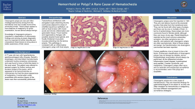
Case Description/Methods: A 74-year-old man underwent colonoscopy for surveillance of polyps. Before the procedure he voiced new-onset painless hematochezia occurring for the last month. A colonoscopy six years earlier had revealed multiple tubular adenomas in addition to small internal hemorrhoids. On his current exam, retroflexed and forward view of the rectum revealed a 1.5cm frond-like lesion atop an internal hemorrhoid near the dentate line prolapsing into the anal canal. Due to concern for an underlying neoplastic process, the patient underwent rectal exam under anesthesia with surgical excision of the lesion.
Histopathology revealed tubulovillous architecture with elongated crypts stretching into the submucosa adjacent areas of stratified squamous epithelium, as well as focal erosion with mixed acute and chronic inflammation and granulation tissue. Reactive and regenerative changes were seen with fused glands with atypia. The muscularis mucosa was focally thickened with extension into the lamina propria. The pathology findings were consistent with an inflammatory cloacogenic polyp with focal atypia.
Discussion: Cloacogenic polyps are rare lesions found at the anorectal junction that arise from the transitional zone, although they may also arise in the colonic lumen. They are thought to occur due to chronic mucosal prolapse but are also associated with Crohns disease and other gastrointestinal disorders. They are more commonly found in adult females but can occur at any age. While often asymptomatic, rectal bleeding is reported as the most common presenting symptom. They are benign, but transformation into cloacogenic carcinomas has been reported. Endoscopic classification is not reliable so histopathologic examination should always be performed. Cloacogenic polyps often recur, so surveillance is recommended after endoscopic or surgical resection, even if dysplasia is not seen. The cloacogenic polyp is a rare, albeit important, finding to identify for gastroenterologists, and should be considered when encountering a polypoid anorectal lesion.

Disclosures:
Michael A. Perrin, MD, MPH, Linda Green, MD, Rollin George, MD. P3151 - Inflammatory Cloacogenic Polyp: An Uncommon Cause of Rectal Outlet Bleeding, ACG 2023 Annual Scientific Meeting Abstracts. Vancouver, BC, Canada: American College of Gastroenterology.
Baylor College of Medicine, Houston, TX
Introduction: Colonoscopy is often ordered to evaluate rectal outlet bleeding and requires careful visual and endoscopic evaluation of the anorectum. Failure to do so may lead to delay in diagnosis or missed premalignant lesions. Here we discuss an uncommon cause of rectal outlet bleeding encountered during surveillance colonoscopy.
Case Description/Methods: A 74-year-old man underwent colonoscopy for surveillance of polyps. Before the procedure he voiced new-onset painless hematochezia occurring for the last month. A colonoscopy six years earlier had revealed multiple tubular adenomas in addition to small internal hemorrhoids. On his current exam, retroflexed and forward view of the rectum revealed a 1.5cm frond-like lesion atop an internal hemorrhoid near the dentate line prolapsing into the anal canal. Due to concern for an underlying neoplastic process, the patient underwent rectal exam under anesthesia with surgical excision of the lesion.
Histopathology revealed tubulovillous architecture with elongated crypts stretching into the submucosa adjacent areas of stratified squamous epithelium, as well as focal erosion with mixed acute and chronic inflammation and granulation tissue. Reactive and regenerative changes were seen with fused glands with atypia. The muscularis mucosa was focally thickened with extension into the lamina propria. The pathology findings were consistent with an inflammatory cloacogenic polyp with focal atypia.
Discussion: Cloacogenic polyps are rare lesions found at the anorectal junction that arise from the transitional zone, although they may also arise in the colonic lumen. They are thought to occur due to chronic mucosal prolapse but are also associated with Crohns disease and other gastrointestinal disorders. They are more commonly found in adult females but can occur at any age. While often asymptomatic, rectal bleeding is reported as the most common presenting symptom. They are benign, but transformation into cloacogenic carcinomas has been reported. Endoscopic classification is not reliable so histopathologic examination should always be performed. Cloacogenic polyps often recur, so surveillance is recommended after endoscopic or surgical resection, even if dysplasia is not seen. The cloacogenic polyp is a rare, albeit important, finding to identify for gastroenterologists, and should be considered when encountering a polypoid anorectal lesion.

Figure: Frond-like polypoid lesion seen at the denate line (Image A) and prolapsing through the anal canal (Image B)
Disclosures:
Michael Perrin indicated no relevant financial relationships.
Linda Green indicated no relevant financial relationships.
Rollin George indicated no relevant financial relationships.
Michael A. Perrin, MD, MPH, Linda Green, MD, Rollin George, MD. P3151 - Inflammatory Cloacogenic Polyp: An Uncommon Cause of Rectal Outlet Bleeding, ACG 2023 Annual Scientific Meeting Abstracts. Vancouver, BC, Canada: American College of Gastroenterology.
