Tuesday Poster Session
Category: Small Intestine
P4168 - Asymptomatic Recurrent Idiopathic Pneumoperitoneum: A Rare Case Presentation
Tuesday, October 24, 2023
10:30 AM - 4:00 PM PT
Location: Exhibit Hall

Has Audio

Mary Vetter, ANPC
University of Rochester Medical Center
Rochester, NY
Presenting Author(s)
Mary Vetter, ANPC1, Sarah Enslin, PA-C1, Victoria Howard, PA-C1, Michael Colicchio, PA-C2, Michael J. Nabozny, MD3, Patrick Fultz, MD1, Derrick German, MD1, Shivangi Kothari, MD3
1University of Rochester Medical Center, Rochester, NY; 2URMC, Rochester, NY; 3University of Rochester, Rochester, NY
Introduction: Abdominal pneumoperitoneum is often caused by perforated abdominal viscus. Other causes include bowel obstruction, appendicitis, neoplasm, vasculitis, and infection. Presentation varies from asymptomatic to life-threatening sepsis and may warrant emergent surgical intervention. Recurrent idiopathic pneumoperitoneum is a very rare disorder described in only a few case reports in literature. We present a case of asymptomatic recurrent idiopathic pneumoperitoneum.
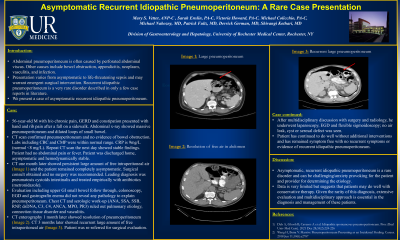
Case Description/Methods: 56-year-old M with h/o chronic pain, GERD and constipation presented with hand and rib pain after a fall on a sidewalk. Abdominal x-ray showed massive pneumoperitoneum and dilated loops of small bowel. CT scan confirmed pneumoperitoneum and no evidence of bowel obstruction. Labs including CBC and CMP were within normal range. CRP was 9mg/L (normal < 8 mg/L). Repeat CT scan the next day showed stable findings. Patient had no abdominal pain or fever. Patient was discharged home, asymptomatic and hemodynamically stable. CT one month later showed persistent large amount of free intraperitoneal air (Image 1). Patient remained completely asymptomatic. Surgical consult obtained and no surgery was recommended. Leading diagnosis was pneumatosis cystoids intestinalis and patient treated empirically with antibiotics (metronidazole). Evaluation including upper GI small bowel follow through, colonoscopy, EGD and gastrograffin enema did not reveal any pathology to explain pneumoperitoneum. Chest CT and serologic work-up (ANA, SSA, SSB, RNP, dsDNA, C3, C4, ANCA, MPO, PR3) ruled out pulmonary etiology, connection tissue disorder and vasculitis. CT enterography 1 month later showed resolution of pneumoperitoneum (Image 2). CT 3 months later showed recurrent large amount of free intraperitoneal air (Image 3). Patient was re-referred for surgical evaluation. After multidisciplinary discussion with surgery and radiology, he underwent laparoscopy, EGD and flexible sigmoidoscopy; no air leak, cyst or serosal defect was seen. Patient has continued to do well without additional intervention.
Discussion: Asymptomatic, recurrent idiopathic pneumoperitoneum is a rare disorder and identifying the etiology can be challenging/anxiety provoking for the patient and provider. Data is very limited but suggests that patients may do well with conservative therapy. Given the rarity of this diagnosis, extensive evaluation and multidisciplinary approach is essential in the diagnosis and management of these patients.

Disclosures:
Mary Vetter, ANPC1, Sarah Enslin, PA-C1, Victoria Howard, PA-C1, Michael Colicchio, PA-C2, Michael J. Nabozny, MD3, Patrick Fultz, MD1, Derrick German, MD1, Shivangi Kothari, MD3. P4168 - Asymptomatic Recurrent Idiopathic Pneumoperitoneum: A Rare Case Presentation, ACG 2023 Annual Scientific Meeting Abstracts. Vancouver, BC, Canada: American College of Gastroenterology.
1University of Rochester Medical Center, Rochester, NY; 2URMC, Rochester, NY; 3University of Rochester, Rochester, NY
Introduction: Abdominal pneumoperitoneum is often caused by perforated abdominal viscus. Other causes include bowel obstruction, appendicitis, neoplasm, vasculitis, and infection. Presentation varies from asymptomatic to life-threatening sepsis and may warrant emergent surgical intervention. Recurrent idiopathic pneumoperitoneum is a very rare disorder described in only a few case reports in literature. We present a case of asymptomatic recurrent idiopathic pneumoperitoneum.
Case Description/Methods: 56-year-old M with h/o chronic pain, GERD and constipation presented with hand and rib pain after a fall on a sidewalk. Abdominal x-ray showed massive pneumoperitoneum and dilated loops of small bowel. CT scan confirmed pneumoperitoneum and no evidence of bowel obstruction. Labs including CBC and CMP were within normal range. CRP was 9mg/L (normal < 8 mg/L). Repeat CT scan the next day showed stable findings. Patient had no abdominal pain or fever. Patient was discharged home, asymptomatic and hemodynamically stable. CT one month later showed persistent large amount of free intraperitoneal air (Image 1). Patient remained completely asymptomatic. Surgical consult obtained and no surgery was recommended. Leading diagnosis was pneumatosis cystoids intestinalis and patient treated empirically with antibiotics (metronidazole). Evaluation including upper GI small bowel follow through, colonoscopy, EGD and gastrograffin enema did not reveal any pathology to explain pneumoperitoneum. Chest CT and serologic work-up (ANA, SSA, SSB, RNP, dsDNA, C3, C4, ANCA, MPO, PR3) ruled out pulmonary etiology, connection tissue disorder and vasculitis. CT enterography 1 month later showed resolution of pneumoperitoneum (Image 2). CT 3 months later showed recurrent large amount of free intraperitoneal air (Image 3). Patient was re-referred for surgical evaluation. After multidisciplinary discussion with surgery and radiology, he underwent laparoscopy, EGD and flexible sigmoidoscopy; no air leak, cyst or serosal defect was seen. Patient has continued to do well without additional intervention.
Discussion: Asymptomatic, recurrent idiopathic pneumoperitoneum is a rare disorder and identifying the etiology can be challenging/anxiety provoking for the patient and provider. Data is very limited but suggests that patients may do well with conservative therapy. Given the rarity of this diagnosis, extensive evaluation and multidisciplinary approach is essential in the diagnosis and management of these patients.

Figure: Recurrent idiopathic pneumoperitoneum CT scans over the course of time showing interval resolution and then recurrence of free air (marked by red arrow) in the peritoneum.
Disclosures:
Mary Vetter indicated no relevant financial relationships.
Sarah Enslin: Castle Biosciences – Advisor or Review Panel Member. Exact Sciences – Advisor or Review Panel Member. Regeneron Pharmaceuticals – Advisor or Review Panel Member.
Victoria Howard indicated no relevant financial relationships.
Michael Colicchio indicated no relevant financial relationships.
Michael Nabozny indicated no relevant financial relationships.
Patrick Fultz indicated no relevant financial relationships.
Derrick German indicated no relevant financial relationships.
Shivangi Kothari: Boston scientific – Consultant. Castle Biosciences – Advisory Committee/Board Member. Olympus – Consultant.
Mary Vetter, ANPC1, Sarah Enslin, PA-C1, Victoria Howard, PA-C1, Michael Colicchio, PA-C2, Michael J. Nabozny, MD3, Patrick Fultz, MD1, Derrick German, MD1, Shivangi Kothari, MD3. P4168 - Asymptomatic Recurrent Idiopathic Pneumoperitoneum: A Rare Case Presentation, ACG 2023 Annual Scientific Meeting Abstracts. Vancouver, BC, Canada: American College of Gastroenterology.
