Tuesday Poster Session
Category: Small Intestine
P4169 - What’s the Leading Point? A Benign Polypoid Mass Behind the Trouble
Tuesday, October 24, 2023
10:30 AM - 4:00 PM PT
Location: Exhibit Hall

Has Audio

Aastha V. Bharwad, MD
University of Kansas School of Medicine
Wichita, KS
Presenting Author(s)
Aastha Bharwad, MD, Chase Branstetter, MD, Mahmoud Mahdi, MD, Nathan D. Tofteland, MD
University of Kansas School of Medicine, Wichita, KS
Introduction: In adult intussusception, an organic lesion has often been shown to act as a leading point. Benign organic lesions are frequent in entero-enteric locations and treatment usually comprises surgical resection of the involved bowel segment.
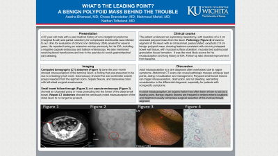
Case Description/Methods: A 67-year-old male with past medical history of non-Hodgkin’s lymphoma (marginal B-cell) and partial colectomy for complicated diverticulitis was referred for evaluation of chronic iron deficiency anemia (IDA) present for seven years. He reported having an extensive workup previously for his IDA, including a negative capsule endoscopy and balloon enteroscopy. He had received blood transfusions and iron in the past due to occult gastrointestinal (GI) bleeding. Computed tomography (CT) abdomen (Figure 1) done the prior month showed intussusception of the terminal ileum, a finding that was presumed to be due to a leading lymph node. Colonoscopy showed five sub-centimeter sessile polyps resected from the sigmoid colon, hepatic flexure, and transverse colon with left-sided surgical anastomosis.
Small bowel follow-through (Figure 2) and capsule endoscopy (Figure 3) showed an ulcerated polyp or mass protruding into the lumen of distal small bowel. CT Abdomen showed the previously noted intussusception in the distal ileum to no longer be present. The patient underwent an exploratory laparotomy, with resection of a 3 cm ulcerated polypoid mass from the ileum.
Pathology (Figure 4) showed a segment of the ileum with an intraluminal, pedunculated, exophytic 2.9 cm benign polypoid mass, showing features consistent with chronic prolapsed bowel wall tissue, with mucosal surface ulceration, mucosal and submucosal granulation tissue formation. It was the most likely source for his intussusception and long history of IDA. Follow-up labs showed improvement from baseline.
Discussion: Adult intussusception is a rare and challenging diagnosis in patients with anemia and/ or obstruction. Diagnosis is often missed because of nonspecific symptoms. In most cases, a pathologic mass acting as a leading point can be identified with abdominal CT that shows the presence of a lead point and can help determine the mass location to guide further management. Polypoid lesions in the small bowel usually present as intussusception or obstruction and have been shown to ulcerate and cause GI bleeding. It is thus important to keep in mind benign polypoid mass in the differential diagnosis of lesions causing intussusception and in those with nonspecific symptoms.

Disclosures:
Aastha Bharwad, MD, Chase Branstetter, MD, Mahmoud Mahdi, MD, Nathan D. Tofteland, MD. P4169 - What’s the Leading Point? A Benign Polypoid Mass Behind the Trouble, ACG 2023 Annual Scientific Meeting Abstracts. Vancouver, BC, Canada: American College of Gastroenterology.
University of Kansas School of Medicine, Wichita, KS
Introduction: In adult intussusception, an organic lesion has often been shown to act as a leading point. Benign organic lesions are frequent in entero-enteric locations and treatment usually comprises surgical resection of the involved bowel segment.
Case Description/Methods: A 67-year-old male with past medical history of non-Hodgkin’s lymphoma (marginal B-cell) and partial colectomy for complicated diverticulitis was referred for evaluation of chronic iron deficiency anemia (IDA) present for seven years. He reported having an extensive workup previously for his IDA, including a negative capsule endoscopy and balloon enteroscopy. He had received blood transfusions and iron in the past due to occult gastrointestinal (GI) bleeding. Computed tomography (CT) abdomen (Figure 1) done the prior month showed intussusception of the terminal ileum, a finding that was presumed to be due to a leading lymph node. Colonoscopy showed five sub-centimeter sessile polyps resected from the sigmoid colon, hepatic flexure, and transverse colon with left-sided surgical anastomosis.
Small bowel follow-through (Figure 2) and capsule endoscopy (Figure 3) showed an ulcerated polyp or mass protruding into the lumen of distal small bowel. CT Abdomen showed the previously noted intussusception in the distal ileum to no longer be present. The patient underwent an exploratory laparotomy, with resection of a 3 cm ulcerated polypoid mass from the ileum.
Pathology (Figure 4) showed a segment of the ileum with an intraluminal, pedunculated, exophytic 2.9 cm benign polypoid mass, showing features consistent with chronic prolapsed bowel wall tissue, with mucosal surface ulceration, mucosal and submucosal granulation tissue formation. It was the most likely source for his intussusception and long history of IDA. Follow-up labs showed improvement from baseline.
Discussion: Adult intussusception is a rare and challenging diagnosis in patients with anemia and/ or obstruction. Diagnosis is often missed because of nonspecific symptoms. In most cases, a pathologic mass acting as a leading point can be identified with abdominal CT that shows the presence of a lead point and can help determine the mass location to guide further management. Polypoid lesions in the small bowel usually present as intussusception or obstruction and have been shown to ulcerate and cause GI bleeding. It is thus important to keep in mind benign polypoid mass in the differential diagnosis of lesions causing intussusception and in those with nonspecific symptoms.

Figure: Figure 1: CT abdomen: Intussusception (arrow) of the terminal ileum
Figure 2: Small bowel follow-through: Area of contrast filling defect (circled)
Figure 3: Capsule endoscopy: Ulcerated polyp or mass (arrow) protruding into the lumen of distal small bowel
Figure 4: Pathology: Benign polypoid mass, with lesion showing features of marked fibromuscular proliferation in the submucosa, thickened muscularis propria, prolapse-type changes, and features of chronic mucosal injury.
Disclosures:
Aastha Bharwad indicated no relevant financial relationships.
Chase Branstetter indicated no relevant financial relationships.
Mahmoud Mahdi indicated no relevant financial relationships.
Nathan Tofteland indicated no relevant financial relationships.
Aastha Bharwad, MD, Chase Branstetter, MD, Mahmoud Mahdi, MD, Nathan D. Tofteland, MD. P4169 - What’s the Leading Point? A Benign Polypoid Mass Behind the Trouble, ACG 2023 Annual Scientific Meeting Abstracts. Vancouver, BC, Canada: American College of Gastroenterology.
