Tuesday Poster Session
Category: Stomach
P4197 - A Scary but Rarely Hazardous Finding
Tuesday, October 24, 2023
10:30 AM - 4:00 PM PT
Location: Exhibit Hall

Has Audio

Spyridon Zouridis, MD
Albany Medical Center
Albany, NY
Presenting Author(s)
Spyridon Zouridis, MD, Omar Tageldin, MD, Joseph Polito, MD
Albany Medical Center, Albany, NY
Introduction: The presence of air within the gastric wall is a rare and worrisome finding. Several conditions leading to gastric intramural air can be lethal if left untreated, however, most conditions that lead to the introduction of air into the gastric wall are benign. This is a case of gastric pneumatosis which was successfully managed with conservative measures.
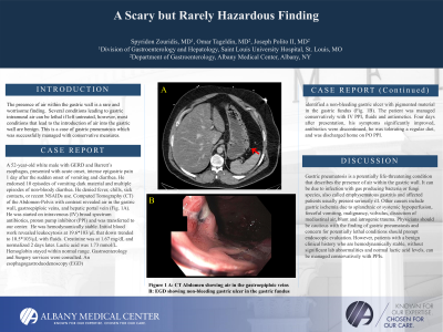
Case Description/Methods: A 52-year-old white male with GERD and Barrett’s esophagus, presented with acute onset, intense epigastric pain 1 day after the sudden onset of vomiting and diarrhea. He endorsed 10 episodes of vomiting dark material and multiple episodes of non-bloody diarrhea. He denied fever, chills, sick contacts, or recent non-steroidal anti-inflammatory drugs use. Computed Tomography (CT) of the Abdomen-Pelvis with contrast revealed air in the gastric wall, gastroepiploic veins, and hepatic portal vein (Fig. A). He was started on intravenous (IV) broad spectrum antibiotics, proton pump inhibitor (PPI) and was transferred to our center. He was hemodynamically stable. Initial blood work revealed leukocytosis at 19.6*103/μL that down trended to 10.5*103/μL with fluids. Creatinine was at 1.67 mg/dL and normalized 2 days later. Lactic acid was 1.73 mmol/L. Hemoglobin stayed within normal range. Gastroenterology and Surgery services were consulted. An esophagogastroduodenoscopy (EGD) identified a non-bleeding gastric ulcer with pigmented material in the gastric fundus (Fig. B). The patient was managed conservatively with IV PPI, fluids and antiemetics. Four days after presentation, his symptoms significantly improved, antibiotics were discontinued, he was tolerating a regular diet, and was discharged home on per os PPI.
Discussion: Gastric pneumatosis is a potentially life-threatening condition that describes the presence of air within the gastric wall. It can be due to infection with gas producing bacteria or fungi species, also called emphysematous gastritis and affected patients usually present seriously ill. Other causes include gastric ischemia due to splanchnic or systemic hypoperfusion, forceful vomiting, malignancy, volvulus, dissection of mediastinal air, blunt and iatrogenic trauma. Physicians should be cautious with the finding of gastric pneumatosis and concern for potentially lethal conditions should prompt endoscopic evaluation. However, patients with a benign clinical history who are hemodynamically stable, without significant lab abnormalities and normal lactic acid levels, can be managed conservatively with PPIs.

Disclosures:
Spyridon Zouridis, MD, Omar Tageldin, MD, Joseph Polito, MD. P4197 - A Scary but Rarely Hazardous Finding, ACG 2023 Annual Scientific Meeting Abstracts. Vancouver, BC, Canada: American College of Gastroenterology.
Albany Medical Center, Albany, NY
Introduction: The presence of air within the gastric wall is a rare and worrisome finding. Several conditions leading to gastric intramural air can be lethal if left untreated, however, most conditions that lead to the introduction of air into the gastric wall are benign. This is a case of gastric pneumatosis which was successfully managed with conservative measures.
Case Description/Methods: A 52-year-old white male with GERD and Barrett’s esophagus, presented with acute onset, intense epigastric pain 1 day after the sudden onset of vomiting and diarrhea. He endorsed 10 episodes of vomiting dark material and multiple episodes of non-bloody diarrhea. He denied fever, chills, sick contacts, or recent non-steroidal anti-inflammatory drugs use. Computed Tomography (CT) of the Abdomen-Pelvis with contrast revealed air in the gastric wall, gastroepiploic veins, and hepatic portal vein (Fig. A). He was started on intravenous (IV) broad spectrum antibiotics, proton pump inhibitor (PPI) and was transferred to our center. He was hemodynamically stable. Initial blood work revealed leukocytosis at 19.6*103/μL that down trended to 10.5*103/μL with fluids. Creatinine was at 1.67 mg/dL and normalized 2 days later. Lactic acid was 1.73 mmol/L. Hemoglobin stayed within normal range. Gastroenterology and Surgery services were consulted. An esophagogastroduodenoscopy (EGD) identified a non-bleeding gastric ulcer with pigmented material in the gastric fundus (Fig. B). The patient was managed conservatively with IV PPI, fluids and antiemetics. Four days after presentation, his symptoms significantly improved, antibiotics were discontinued, he was tolerating a regular diet, and was discharged home on per os PPI.
Discussion: Gastric pneumatosis is a potentially life-threatening condition that describes the presence of air within the gastric wall. It can be due to infection with gas producing bacteria or fungi species, also called emphysematous gastritis and affected patients usually present seriously ill. Other causes include gastric ischemia due to splanchnic or systemic hypoperfusion, forceful vomiting, malignancy, volvulus, dissection of mediastinal air, blunt and iatrogenic trauma. Physicians should be cautious with the finding of gastric pneumatosis and concern for potentially lethal conditions should prompt endoscopic evaluation. However, patients with a benign clinical history who are hemodynamically stable, without significant lab abnormalities and normal lactic acid levels, can be managed conservatively with PPIs.

Figure: A: Computed Tomography Abdomen Pelvis (orange arrow: air in gastroepiploic veins)
B: Esophagogastroduodenoscopy showing a non-bleeding gastric ulcer with pigmented material in the gastric fundus
B: Esophagogastroduodenoscopy showing a non-bleeding gastric ulcer with pigmented material in the gastric fundus
Disclosures:
Spyridon Zouridis indicated no relevant financial relationships.
Omar Tageldin indicated no relevant financial relationships.
Joseph Polito indicated no relevant financial relationships.
Spyridon Zouridis, MD, Omar Tageldin, MD, Joseph Polito, MD. P4197 - A Scary but Rarely Hazardous Finding, ACG 2023 Annual Scientific Meeting Abstracts. Vancouver, BC, Canada: American College of Gastroenterology.
