Tuesday Poster Session
Category: Stomach
P4203 - She Ate What?! Bird Seeds & Cat Food - A Case of Rectal & Gastric Bezoar
Tuesday, October 24, 2023
10:30 AM - 4:00 PM PT
Location: Exhibit Hall

Has Audio
- HN
Haider Abbas A. Naqvi, MD
MedStar Health
Baltimore, MD
Presenting Author(s)
Joseph O. Atarere, MBChB, MPH1, Haider A. Naqvi, MD2
1MedStar Union Memorial Hospital, Baltimore, MD; 2MedStar Health, Baltimore, MD
Introduction: Most cases of gastrointestinal (GI) bezoars occur in the stomach and small bowel. Phytobezoars are the most common type of GI bezoars (~40% of cases). Seed bezoars are a subcategory of phytobezoars responsible for >50% of cases of fecal impaction requiring hospitalization but are a rare cause of gastric bezoars.
Case Description/Methods: A wheelchair bound 48-year-old woman with cognitive developmental delay was admitted to the hospital with progressively worsening constipation followed by 1 week of diarrhea and 1 day of periumbilical and rectal pain. Due to financial and mobility difficulties, she ran out of food and for several months, her diet consisted of cat food and bird seeds. On physical exam she was cachetic with abdominal distention and mild tenderness. Numerous sunflower seeds were visible at the anal verge on rectal inspection. CT scan of the abdomen and pelvis showed massive stool burden throughout the GI tract with dilated rectum and rectal wall thickening and surrounding edema. She was taken to the operating room for manual evacuation under anesthesia with findings of liquid stool, seeds and a pieces of corn. Post-operative CT scan of the abdomen and pelvis with oral contrast revealed a completely decompressed rectum, normal caliber transverse and left hemicolon with moderate fecal material in the right hemicolon and terminal ileum She was subsequently managed with laxatives with passage of moderate amounts of liquid stool. Repeat scans on postoperative day 5 showed marked gastric dilation with increased stool burden in the colon. A diagnosis of gastric bezoar was made and treatment with Coca-Cola was initiated. CT abdomen and pelvis on postoperative day 10 showed resolution of gastric distension with liquid stool throughout the colon and she was discharged to rehab with standing bowel regimen.
Discussion: A diagnosis of GI bezoar is often suggested by a typical history and rectal exam. CT scan is the most reliable modality for diagnosing GI bezoars but endoscopy is the gold standard for diagnosing gastric bezoars. Rectal seed bezoars are best managed with manual evacuation under anesthesia. Management of gastric phytobezoars can be divided into: dissolution, fragmentation, and/or retrieval. In the case described above, the bezoar resolved completely with dissolution therapy using Coca-Cola. Fragmentation and retrieval by endoscopy is reserved for cases unresponsive to dissolution therapy.

Disclosures:
Joseph O. Atarere, MBChB, MPH1, Haider A. Naqvi, MD2. P4203 - She Ate What?! Bird Seeds & Cat Food - A Case of Rectal & Gastric Bezoar, ACG 2023 Annual Scientific Meeting Abstracts. Vancouver, BC, Canada: American College of Gastroenterology.
1MedStar Union Memorial Hospital, Baltimore, MD; 2MedStar Health, Baltimore, MD
Introduction: Most cases of gastrointestinal (GI) bezoars occur in the stomach and small bowel. Phytobezoars are the most common type of GI bezoars (~40% of cases). Seed bezoars are a subcategory of phytobezoars responsible for >50% of cases of fecal impaction requiring hospitalization but are a rare cause of gastric bezoars.
Case Description/Methods: A wheelchair bound 48-year-old woman with cognitive developmental delay was admitted to the hospital with progressively worsening constipation followed by 1 week of diarrhea and 1 day of periumbilical and rectal pain. Due to financial and mobility difficulties, she ran out of food and for several months, her diet consisted of cat food and bird seeds. On physical exam she was cachetic with abdominal distention and mild tenderness. Numerous sunflower seeds were visible at the anal verge on rectal inspection. CT scan of the abdomen and pelvis showed massive stool burden throughout the GI tract with dilated rectum and rectal wall thickening and surrounding edema. She was taken to the operating room for manual evacuation under anesthesia with findings of liquid stool, seeds and a pieces of corn. Post-operative CT scan of the abdomen and pelvis with oral contrast revealed a completely decompressed rectum, normal caliber transverse and left hemicolon with moderate fecal material in the right hemicolon and terminal ileum She was subsequently managed with laxatives with passage of moderate amounts of liquid stool. Repeat scans on postoperative day 5 showed marked gastric dilation with increased stool burden in the colon. A diagnosis of gastric bezoar was made and treatment with Coca-Cola was initiated. CT abdomen and pelvis on postoperative day 10 showed resolution of gastric distension with liquid stool throughout the colon and she was discharged to rehab with standing bowel regimen.
Discussion: A diagnosis of GI bezoar is often suggested by a typical history and rectal exam. CT scan is the most reliable modality for diagnosing GI bezoars but endoscopy is the gold standard for diagnosing gastric bezoars. Rectal seed bezoars are best managed with manual evacuation under anesthesia. Management of gastric phytobezoars can be divided into: dissolution, fragmentation, and/or retrieval. In the case described above, the bezoar resolved completely with dissolution therapy using Coca-Cola. Fragmentation and retrieval by endoscopy is reserved for cases unresponsive to dissolution therapy.

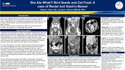
Figure: Panels A&B: CT scan of the abdomen and pelvis on initial presentation showing the dilated rectum, rectal wall thickening with surrounding edema and rectal bezoar.
Panels C&D: CT scan of the abdomen and pelvis after manual evacuation under anesthesia showing a normal caliber transverse and left hemicolon with moderate fecal material in the right hemicolon (C:axial, D: coronal view).
Panel E: CT scan of the abdomen and pelvis on postoperative day 5 showing marked gastric dilation and gastric bezoar with increased stool burden in the colon
Panel F: CT scan of the abdomen and pelvis on postoperative day 10 showing resolution of the gastric distension
Disclosures:
Joseph Atarere indicated no relevant financial relationships.
Haider Naqvi indicated no relevant financial relationships.
Joseph O. Atarere, MBChB, MPH1, Haider A. Naqvi, MD2. P4203 - She Ate What?! Bird Seeds & Cat Food - A Case of Rectal & Gastric Bezoar, ACG 2023 Annual Scientific Meeting Abstracts. Vancouver, BC, Canada: American College of Gastroenterology.
