Tuesday Poster Session
Category: Stomach
P4204 - Metastatic Melanoma Presents as Hypopigmented Gastric Nodules
Tuesday, October 24, 2023
10:30 AM - 4:00 PM PT
Location: Exhibit Hall

Has Audio

Nicole L. Miller, MD
University of South Florida Morsani College of Medicine
Tampa, FL
Presenting Author(s)
Nicole L. Miller, MD, Kevin Shah, MD, Rene Gomez Esquivel, MD
University of South Florida Morsani College of Medicine, Tampa, FL
Introduction: Metastatic melanoma to the gastrointestinal tract often occurs in the small intestine and colon with rare involvement of the stomach1. Endoscopic features of metastatic melanoma include small nodules, black spots, and ulcers. This case demonstrates an unusual presentation of melanoma metastases presenting as multiple gastric polyps with hypopigmentation resembling amelanotic melanoma.
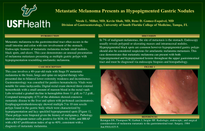
Case Description/Methods: This case involves a 40-year-old male with Stage IV metastatic melanoma to the brain, lungs and spine on targeted therapy who presented due to bilateral lower extremity weakness and incontinence. Gastroenterology was consulted for painless hematochezia. Vitals were notable for sinus tachycardia. Digital rectal exam showed three external hemorrhoids with a small amount of maroon blood in the rectal vault. Labs revealed a gradual decline in hemoglobin from 11 g/dL to 7.2 g/dL. Computed tomography (CT) of the abdomen showed extensive metastatic disease to the liver and spleen with peritoneal carcinomatosis. Esophagogastroduodenoscopy showed multiple 5 to 10 mm sessile polyps in the gastric body characterized by hypopigmentation and lacy speckled hyperpigmented spots [Figure 1]. These polyps were biopsied given the history of malignancy. Pathology showed malignant tumor cells positive for SOX-10, S100, and BRAF with a KI 67 proliferation index of up to 40%, consistent with a diagnosis of metastatic melanoma. Due to cord compression and neurological decline, the patient transitioned to hospice care and passed away one month later.
Discussion: In 7% of malignant melanomas, the site of metastasis is the stomach. Endoscopy usually reveals polypoid or ulcerating masses and intramucosal nodules1. Hyperpigmented black spots are common however hypopigmented gastric polyps should also be considered suspicious for amelanotic melanoma metastasis. This case demonstrates that metastatic melanoma can present with both hyperpigmented and hypopigmented lesions throughout the upper gastrointestinal tract and must be diagnosed via endoscopic biopsies and histopathology.
References:
1)Reintgen, et al. Radiologic, endoscopic, and surgical considerations of melanoma metastatic to the gastrointestinal tract. Surgery. June 1984.

Disclosures:
Nicole L. Miller, MD, Kevin Shah, MD, Rene Gomez Esquivel, MD. P4204 - Metastatic Melanoma Presents as Hypopigmented Gastric Nodules, ACG 2023 Annual Scientific Meeting Abstracts. Vancouver, BC, Canada: American College of Gastroenterology.
University of South Florida Morsani College of Medicine, Tampa, FL
Introduction: Metastatic melanoma to the gastrointestinal tract often occurs in the small intestine and colon with rare involvement of the stomach1. Endoscopic features of metastatic melanoma include small nodules, black spots, and ulcers. This case demonstrates an unusual presentation of melanoma metastases presenting as multiple gastric polyps with hypopigmentation resembling amelanotic melanoma.
Case Description/Methods: This case involves a 40-year-old male with Stage IV metastatic melanoma to the brain, lungs and spine on targeted therapy who presented due to bilateral lower extremity weakness and incontinence. Gastroenterology was consulted for painless hematochezia. Vitals were notable for sinus tachycardia. Digital rectal exam showed three external hemorrhoids with a small amount of maroon blood in the rectal vault. Labs revealed a gradual decline in hemoglobin from 11 g/dL to 7.2 g/dL. Computed tomography (CT) of the abdomen showed extensive metastatic disease to the liver and spleen with peritoneal carcinomatosis. Esophagogastroduodenoscopy showed multiple 5 to 10 mm sessile polyps in the gastric body characterized by hypopigmentation and lacy speckled hyperpigmented spots [Figure 1]. These polyps were biopsied given the history of malignancy. Pathology showed malignant tumor cells positive for SOX-10, S100, and BRAF with a KI 67 proliferation index of up to 40%, consistent with a diagnosis of metastatic melanoma. Due to cord compression and neurological decline, the patient transitioned to hospice care and passed away one month later.
Discussion: In 7% of malignant melanomas, the site of metastasis is the stomach. Endoscopy usually reveals polypoid or ulcerating masses and intramucosal nodules1. Hyperpigmented black spots are common however hypopigmented gastric polyps should also be considered suspicious for amelanotic melanoma metastasis. This case demonstrates that metastatic melanoma can present with both hyperpigmented and hypopigmented lesions throughout the upper gastrointestinal tract and must be diagnosed via endoscopic biopsies and histopathology.
References:
1)Reintgen, et al. Radiologic, endoscopic, and surgical considerations of melanoma metastatic to the gastrointestinal tract. Surgery. June 1984.

Figure: Figure 1. Hypopigmented gastric metastatic melanoma nodule
Disclosures:
Nicole Miller indicated no relevant financial relationships.
Kevin Shah indicated no relevant financial relationships.
Rene Gomez Esquivel indicated no relevant financial relationships.
Nicole L. Miller, MD, Kevin Shah, MD, Rene Gomez Esquivel, MD. P4204 - Metastatic Melanoma Presents as Hypopigmented Gastric Nodules, ACG 2023 Annual Scientific Meeting Abstracts. Vancouver, BC, Canada: American College of Gastroenterology.

