Tuesday Poster Session
Category: Stomach
P4224 - A Case of Gastric Emphysema Associated With Gastroparesis
Tuesday, October 24, 2023
10:30 AM - 4:00 PM PT
Location: Exhibit Hall

Has Audio

Noor Siddiqi Syed, MD
Santa Clara Valley Medical Center
Saratoga, CA
Presenting Author(s)
Noor Syed, MD1, Scott Ventre, DO2, Anish V. Patel, MD3
1Santa Clara Valley Medical Center, Saratoga, CA; 2Robert Wood Johnson University Hospital, New Brunswick, NJ; 3Rutgers Robert Wood Johnson Medical School, New Brunswick, NJ
Introduction: Gastric emphysema is a radiographic finding defined by the presence of air within the walls of the stomach. It can be benign and self limiting but also due to infection, ischemia or related to a gastric outlet obstruction from masses or strictures. Often clinicians face a dilemma of pursuing conservative management or surgical interventions.
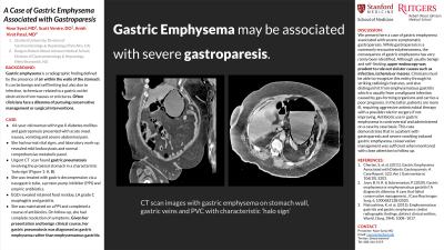
Case Description/Methods: A 64-year-old woman with type II diabetes mellitus and gastroparesis presented with acute onset nausea, vomiting and abdominal pain. Laboratory work-up revealed mild leukocytosis and normal comprehensive metabolic panel. On presentation, she had normal vital signs, but was noted to have diffuse abdominal tenderness. Urgent computerized tomography scan found gastric pneumatosis involving the proximal stomach in a characteristic ‘halo sign’ (Figure 1: A, B). Gastric decompression with a nasal-gastric tube provided significant symptom relief and she was started on a proton-pump inhibitor (PPI) and empiric antibiotics. Upper endoscopy revealed significant retained food residue, LA grade C esophagitis and mild gastritis. She was administered metoclopramide and her glycemic control was optimized. She was maintained on PPI therapy and completed a course of antibiotics. On follow-up, she had complete resolution of symptoms. Given her presentation and benign clinical course, her gastric pneumatosis was diagnosed as gastric emphysema rather than emphysematous gastritis.
Discussion: We present here a case of gastric emphysema associated with severe symptomatic gastroparesis. While gastroparesis is a commonly encountered phenomena, the consequence of gastric emphysema has very rarely been identified. Although usually benign and self-limiting, upper endoscopy was prudent to rule out sinister causes such as infection, ischemia or masses. Clinicians must be able to recognize this entity through its striking radiologic features, and also distinguish it from emphysematous gastritis which is usually from a malignant infection caused by gas-forming organisms and carries a poor prognosis. In the latter, patients are more ill, requiring aggressive antimicrobial therapy with a possible role for surgery if not improving. Antibiotic use in gastric emphysema is controversial and administered on a case by case basis. This case demonstrates that in a patient with gastroparesis and severe vomiting induced gastric emphysema, conservative management was sufficient when monitored with close attention to follow up.

Disclosures:
Noor Syed, MD1, Scott Ventre, DO2, Anish V. Patel, MD3. P4224 - A Case of Gastric Emphysema Associated With Gastroparesis, ACG 2023 Annual Scientific Meeting Abstracts. Vancouver, BC, Canada: American College of Gastroenterology.
1Santa Clara Valley Medical Center, Saratoga, CA; 2Robert Wood Johnson University Hospital, New Brunswick, NJ; 3Rutgers Robert Wood Johnson Medical School, New Brunswick, NJ
Introduction: Gastric emphysema is a radiographic finding defined by the presence of air within the walls of the stomach. It can be benign and self limiting but also due to infection, ischemia or related to a gastric outlet obstruction from masses or strictures. Often clinicians face a dilemma of pursuing conservative management or surgical interventions.
Case Description/Methods: A 64-year-old woman with type II diabetes mellitus and gastroparesis presented with acute onset nausea, vomiting and abdominal pain. Laboratory work-up revealed mild leukocytosis and normal comprehensive metabolic panel. On presentation, she had normal vital signs, but was noted to have diffuse abdominal tenderness. Urgent computerized tomography scan found gastric pneumatosis involving the proximal stomach in a characteristic ‘halo sign’ (Figure 1: A, B). Gastric decompression with a nasal-gastric tube provided significant symptom relief and she was started on a proton-pump inhibitor (PPI) and empiric antibiotics. Upper endoscopy revealed significant retained food residue, LA grade C esophagitis and mild gastritis. She was administered metoclopramide and her glycemic control was optimized. She was maintained on PPI therapy and completed a course of antibiotics. On follow-up, she had complete resolution of symptoms. Given her presentation and benign clinical course, her gastric pneumatosis was diagnosed as gastric emphysema rather than emphysematous gastritis.
Discussion: We present here a case of gastric emphysema associated with severe symptomatic gastroparesis. While gastroparesis is a commonly encountered phenomena, the consequence of gastric emphysema has very rarely been identified. Although usually benign and self-limiting, upper endoscopy was prudent to rule out sinister causes such as infection, ischemia or masses. Clinicians must be able to recognize this entity through its striking radiologic features, and also distinguish it from emphysematous gastritis which is usually from a malignant infection caused by gas-forming organisms and carries a poor prognosis. In the latter, patients are more ill, requiring aggressive antimicrobial therapy with a possible role for surgery if not improving. Antibiotic use in gastric emphysema is controversial and administered on a case by case basis. This case demonstrates that in a patient with gastroparesis and severe vomiting induced gastric emphysema, conservative management was sufficient when monitored with close attention to follow up.

Figure: Figure 1: A, B: CT scan images with gastric emphysema on stomach wall, gastric veins and PVC.
Disclosures:
Noor Syed indicated no relevant financial relationships.
Scott Ventre indicated no relevant financial relationships.
Anish Patel indicated no relevant financial relationships.
Noor Syed, MD1, Scott Ventre, DO2, Anish V. Patel, MD3. P4224 - A Case of Gastric Emphysema Associated With Gastroparesis, ACG 2023 Annual Scientific Meeting Abstracts. Vancouver, BC, Canada: American College of Gastroenterology.
