Tuesday Poster Session
Category: Stomach
P4225 - Rare Presentation of Gastric Ulcer as Gastric Pneumatosis
Tuesday, October 24, 2023
10:30 AM - 4:00 PM PT
Location: Exhibit Hall


Michael Ladna, MD
University of Florida
Gainesville, FL
Presenting Author(s)
Michael Ladna, MD, John George, MD
University of Florida, Gainesville, FL
Introduction: Gastric pneumatosis is an ominous radiographic finding which was associated with a large gastric ulcer in the following case. It is typically managed conservatively via bowel rest, decompression via NG tube, broad-spectrum IV antibiotics, and IV PPI. Surgical intervention is typically only required for gastric perforation or if lack of response to conservative management.
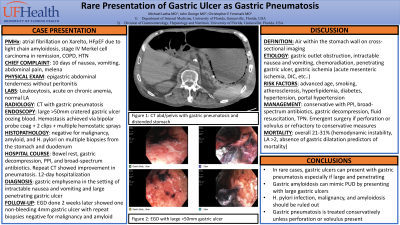
Case Description/Methods: An elderly man with atrial fibrillation on Xarelto, HFpEF due to cardiac amyloidosis, and stage IV Merkel cell carcinoma in remission presented with 10 days of vomiting, abdominal pain, and watery non-bloody diarrhea. CT showed gastric pneumatosis. There was no portal venous gas, bowel obstruction, or pneumoperitoneum. He was made NPO, NG tube was placed, started on piperacillin-tazobactam and vancomycin as well as IV PPI. Surgery was consulted and recommended continuing conservative therapy Repeat CT 3 days later showed interval improvement in gastric pneumatosis. On day 4 he developed an upper GI bleed. Anticoagulation was held and an EGD was done which revealed 1 large >50mm oozing cratered gastric ulcer with an adherent clot in the gastric body. After clot removal, visible vessels were exposed with slow oozing. Hemostasis was achieved via a combination of bipolar probe coagulation, hemostatic clips, and multiple hemostatic sprays. Pathology did not show conclusive evidence of malignancy thus repeat EGD was done 2 weeks later which showed one 4mm non-bleeding gastric ulcer. Pathology showed gastric mucosa with moderate chronic inactive gastritis without any metaplasia, dysplasia, or carcinoma.
Discussion: Gastric pneumatosis is an uncommon and ominous radiographic finding. Gastric pneumatosis can be seen in potentially fatal pathologies such as emphysematous gastritis or gastric ischemia and benign pathologies such as gastric emphysema. There are numerous entities associated with gastric pneumatosis such as gastric outlet obstruction, Intractable nausea and vomiting, penetrating gastric ulcer, and gastric ischemia which has been associated with DIC and mesenteric ischemia. Management of gastric pneumatosis is divided into surgical and conservative. Conservative approaches involve gastric acid suppression with PPI, broad-spectrum antibiotics, nasogastric tube decompression, fluid resuscitation, and TPN. Surgical intervention is recommended for gastric perforation and gangrenous or necrotizing gastritis that does not respond to conservative management or ischemia secondary to gastric volvulus.

Disclosures:
Michael Ladna, MD, John George, MD. P4225 - Rare Presentation of Gastric Ulcer as Gastric Pneumatosis, ACG 2023 Annual Scientific Meeting Abstracts. Vancouver, BC, Canada: American College of Gastroenterology.
University of Florida, Gainesville, FL
Introduction: Gastric pneumatosis is an ominous radiographic finding which was associated with a large gastric ulcer in the following case. It is typically managed conservatively via bowel rest, decompression via NG tube, broad-spectrum IV antibiotics, and IV PPI. Surgical intervention is typically only required for gastric perforation or if lack of response to conservative management.
Case Description/Methods: An elderly man with atrial fibrillation on Xarelto, HFpEF due to cardiac amyloidosis, and stage IV Merkel cell carcinoma in remission presented with 10 days of vomiting, abdominal pain, and watery non-bloody diarrhea. CT showed gastric pneumatosis. There was no portal venous gas, bowel obstruction, or pneumoperitoneum. He was made NPO, NG tube was placed, started on piperacillin-tazobactam and vancomycin as well as IV PPI. Surgery was consulted and recommended continuing conservative therapy Repeat CT 3 days later showed interval improvement in gastric pneumatosis. On day 4 he developed an upper GI bleed. Anticoagulation was held and an EGD was done which revealed 1 large >50mm oozing cratered gastric ulcer with an adherent clot in the gastric body. After clot removal, visible vessels were exposed with slow oozing. Hemostasis was achieved via a combination of bipolar probe coagulation, hemostatic clips, and multiple hemostatic sprays. Pathology did not show conclusive evidence of malignancy thus repeat EGD was done 2 weeks later which showed one 4mm non-bleeding gastric ulcer. Pathology showed gastric mucosa with moderate chronic inactive gastritis without any metaplasia, dysplasia, or carcinoma.
Discussion: Gastric pneumatosis is an uncommon and ominous radiographic finding. Gastric pneumatosis can be seen in potentially fatal pathologies such as emphysematous gastritis or gastric ischemia and benign pathologies such as gastric emphysema. There are numerous entities associated with gastric pneumatosis such as gastric outlet obstruction, Intractable nausea and vomiting, penetrating gastric ulcer, and gastric ischemia which has been associated with DIC and mesenteric ischemia. Management of gastric pneumatosis is divided into surgical and conservative. Conservative approaches involve gastric acid suppression with PPI, broad-spectrum antibiotics, nasogastric tube decompression, fluid resuscitation, and TPN. Surgical intervention is recommended for gastric perforation and gangrenous or necrotizing gastritis that does not respond to conservative management or ischemia secondary to gastric volvulus.

Figure: CT of the abdomen and pelvis showing gastric pneumatosis (top image) and Endoscopy images showing large >50mm cratered gastric ulcer
Disclosures:
Michael Ladna indicated no relevant financial relationships.
John George indicated no relevant financial relationships.
Michael Ladna, MD, John George, MD. P4225 - Rare Presentation of Gastric Ulcer as Gastric Pneumatosis, ACG 2023 Annual Scientific Meeting Abstracts. Vancouver, BC, Canada: American College of Gastroenterology.
