Tuesday Poster Session
Category: Stomach
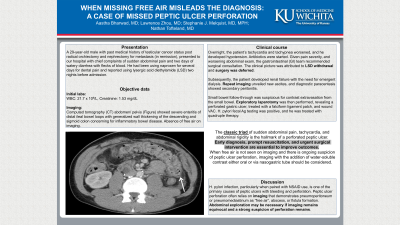
P4247 - When Missing Free Air Misleads the Diagnosis: A Case of Missed Peptic Ulcer Perforation
Tuesday, October 24, 2023
10:30 AM - 4:00 PM PT
Location: Exhibit Hall

Has Audio

Aastha V. Bharwad, MD
University of Kansas School of Medicine
Wichita, KS
Presenting Author(s)
Aastha Bharwad, MD, Lawrence Zhou, MD, Stephanie J. Melquist, MD, MPH, Nathan D. Tofteland, MD
University of Kansas School of Medicine, Wichita, KS
Introduction: The classic triad of sudden abdominal pain, tachycardia, and abdominal rigidity is the hallmark of a perforated peptic ulcer. Early diagnosis, prompt resuscitation, and urgent surgical intervention are essential to improve outcomes. We present a case of a young male who had a delayed diagnosis of peptic ulcer perforation and thus increased morbidity due to the absence of free air on imaging.
Case Description/Methods: A 29-year-old male with a past history of testicular cancer status post radical orchiectomy and nephrectomy for metastasis, in remission, presented to the hospital with sudden abdominal pain that woke him from sleep and two days of watery diarrhea with flecks of blood. He had been using naproxen for several days for dental pain. He also used lysergic acid diethylamide (LSD) two nights before admission. Computed tomography (CT) abdomen/ pelvis (Figure 1) showed severe enteritis of distal ileal bowel loops with generalized wall thickening of descending and sigmoid colon concerning for inflammatory bowel disease. Labs were significant for white blood cell count of 21.7 x 10⁹/ L and creatinine of 1.53 mg/ dL.
Overnight, tachycardia and tachypnea worsened and hypotension developed. Antibiotics were started. Given pain severity, and worsening abdominal exam, the gastrointestinal (GI) team recommended surgical consultation. The clinical picture was attributed to LSD withdrawal and surgery was deferred. Subsequently, he developed renal failure with the need for emergent dialysis. Repeat imaging unveiled new ascites, and diagnostic paracentesis showed secondary peritonitis. Small bowel follow-through was suspicious for contrast extravasation from the small bowel. Exploratory laparotomy was then performed, revealing a perforated gastric ulcer, treated with a falciform ligament patch, and wound VAC. H. pylori fecal Ag testing was positive and he was treated with quadruple therapy.
Discussion: H. pylori infection, particularly when paired with NSAID use, is one of the primary causes of peptic ulcers with bleeding and perforation. Peptic ulcer perforation often relies on imaging that demonstrates pneumoperitoneum or pneumomediastinum as “free air”, abscess, or fistula formation. Abdominal exploration may be necessary if imaging remains equivocal and a strong suspicion of perforation remains. When free air is not seen on imaging and there is ongoing suspicion of perforated peptic ulcer, imaging with the addition of water-soluble contrast either oral or via nasogastric tube should be considered.

Disclosures:
Aastha Bharwad, MD, Lawrence Zhou, MD, Stephanie J. Melquist, MD, MPH, Nathan D. Tofteland, MD. P4247 - When Missing Free Air Misleads the Diagnosis: A Case of Missed Peptic Ulcer Perforation, ACG 2023 Annual Scientific Meeting Abstracts. Vancouver, BC, Canada: American College of Gastroenterology.
University of Kansas School of Medicine, Wichita, KS
Introduction: The classic triad of sudden abdominal pain, tachycardia, and abdominal rigidity is the hallmark of a perforated peptic ulcer. Early diagnosis, prompt resuscitation, and urgent surgical intervention are essential to improve outcomes. We present a case of a young male who had a delayed diagnosis of peptic ulcer perforation and thus increased morbidity due to the absence of free air on imaging.
Case Description/Methods: A 29-year-old male with a past history of testicular cancer status post radical orchiectomy and nephrectomy for metastasis, in remission, presented to the hospital with sudden abdominal pain that woke him from sleep and two days of watery diarrhea with flecks of blood. He had been using naproxen for several days for dental pain. He also used lysergic acid diethylamide (LSD) two nights before admission. Computed tomography (CT) abdomen/ pelvis (Figure 1) showed severe enteritis of distal ileal bowel loops with generalized wall thickening of descending and sigmoid colon concerning for inflammatory bowel disease. Labs were significant for white blood cell count of 21.7 x 10⁹/ L and creatinine of 1.53 mg/ dL.
Overnight, tachycardia and tachypnea worsened and hypotension developed. Antibiotics were started. Given pain severity, and worsening abdominal exam, the gastrointestinal (GI) team recommended surgical consultation. The clinical picture was attributed to LSD withdrawal and surgery was deferred. Subsequently, he developed renal failure with the need for emergent dialysis. Repeat imaging unveiled new ascites, and diagnostic paracentesis showed secondary peritonitis. Small bowel follow-through was suspicious for contrast extravasation from the small bowel. Exploratory laparotomy was then performed, revealing a perforated gastric ulcer, treated with a falciform ligament patch, and wound VAC. H. pylori fecal Ag testing was positive and he was treated with quadruple therapy.
Discussion: H. pylori infection, particularly when paired with NSAID use, is one of the primary causes of peptic ulcers with bleeding and perforation. Peptic ulcer perforation often relies on imaging that demonstrates pneumoperitoneum or pneumomediastinum as “free air”, abscess, or fistula formation. Abdominal exploration may be necessary if imaging remains equivocal and a strong suspicion of perforation remains. When free air is not seen on imaging and there is ongoing suspicion of perforated peptic ulcer, imaging with the addition of water-soluble contrast either oral or via nasogastric tube should be considered.

Figure: Figure 1: CT Abdomen/ Pelvis with contrast: Severe enteritis of the distal ileal bowel loops with reactive free fluid in the abdomen and pelvis. Generalized wall thickening of the descending and sigmoid colon. Underlying IBD is a consideration. Absence of free air on imaging.
Disclosures:
Aastha Bharwad indicated no relevant financial relationships.
Lawrence Zhou indicated no relevant financial relationships.
Stephanie Melquist indicated no relevant financial relationships.
Nathan Tofteland indicated no relevant financial relationships.
Aastha Bharwad, MD, Lawrence Zhou, MD, Stephanie J. Melquist, MD, MPH, Nathan D. Tofteland, MD. P4247 - When Missing Free Air Misleads the Diagnosis: A Case of Missed Peptic Ulcer Perforation, ACG 2023 Annual Scientific Meeting Abstracts. Vancouver, BC, Canada: American College of Gastroenterology.
