Tuesday Poster Session
Category: Stomach
P4257 - Advanced Endoscopic Techniques for a Refractory Pyloric Stricture
Tuesday, October 24, 2023
10:30 AM - 4:00 PM PT
Location: Exhibit Hall

Has Audio

Anastasia Chahine, MD
University of California Irvine
Orange, CA
Presenting Author(s)
Anastasia Chahine, MD, Amirali Tavangar, MD, Jun Ho, , Khaldoon Khirfan, MD, Usman Rahim, MD, Tina Zhou, BS, Jason Samarasena, MD, Kenneth Chang, MD
University of California Irvine, Orange, CA
Introduction: Pyloric strictures can be managed with several endoscopic techniques. Endoscopic balloon dilation, a procedure that uses an inflated balloon to widen the narrowed area, is typically the first-line treatment. Occasionally, a stent is deployed to create continuous dilation of a stricture. Another technique, called Endoscopic Pyloromyotomy, is sometimes used in severe refractory strictures and involves cutting the pyloric muscle to relieve the obstruction. We present here a challenging case of a refractory pyloric stricture where we employed endoscopic stricturoplasty and pyloromyotomy for definitive treatment.
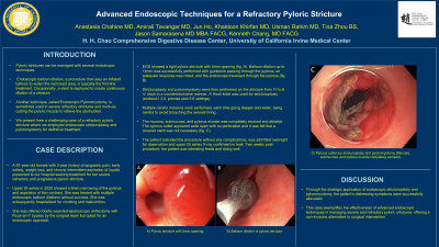
Case Description/Methods: A 67-year-old female with 3-year history of epigastric pain, early satiety, weight loss, and chronic intermittent aspiration of liquids presented to our hospital seeking treatment for her severe refractory and progressive pyloric stricture. Upper GI series showed a 6mm narrowing of the pylorus and aspiration of thin contrast. She was treated with endoscopic balloon dilations without success. She was subsequently hospitalized for vomiting and malnutrition. She was offered laparoscopic antrectomy with Roux-en-Y bypass by the surgical team but opted for an endoscopic approach. EGD showed a tight pyloric stricture with 5mm opening (fig.A). Balloon dilation up to 14mm was successfully performed with guidewire passing through the pylorus, an adequate response was noted, and the endoscope traversed through the pylorus (fig.B). Stricturoplasty and pyloromyotomy were then performed on the stricture from 10 to 6 'o' clock in a counterclockwise manner. A Hook knife was used for stricturoplasty. Multiple careful incisions were performed, each time going deeper and wider, being careful to avoid broaching the serosal lining. The mucosa, submucosa, and pylorus muscle was completely excised and ablated. The pylorus outlet appeared wide open with no perforation and it was felt that a covered stent was not necessary (fig.C). The patient tolerated the procedure without complications, was admitted overnight for observation and upper GI series X-ray confirmed no leak. Two weeks post-procedure, the patient was tolerating feeds and doing well.
Discussion: Through the strategic application of endoscopic stricturoplasty and pyloromyotomy, the patient’s distressing symptoms were successfully alleviated. This case exemplifies the effectiveness of advanced endoscopic techniques in managing severe and refractory pyloric strictures, offering a non-invasive alternative to surgical intervention.

Disclosures:
Anastasia Chahine, MD, Amirali Tavangar, MD, Jun Ho, , Khaldoon Khirfan, MD, Usman Rahim, MD, Tina Zhou, BS, Jason Samarasena, MD, Kenneth Chang, MD. P4257 - Advanced Endoscopic Techniques for a Refractory Pyloric Stricture, ACG 2023 Annual Scientific Meeting Abstracts. Vancouver, BC, Canada: American College of Gastroenterology.
University of California Irvine, Orange, CA
Introduction: Pyloric strictures can be managed with several endoscopic techniques. Endoscopic balloon dilation, a procedure that uses an inflated balloon to widen the narrowed area, is typically the first-line treatment. Occasionally, a stent is deployed to create continuous dilation of a stricture. Another technique, called Endoscopic Pyloromyotomy, is sometimes used in severe refractory strictures and involves cutting the pyloric muscle to relieve the obstruction. We present here a challenging case of a refractory pyloric stricture where we employed endoscopic stricturoplasty and pyloromyotomy for definitive treatment.
Case Description/Methods: A 67-year-old female with 3-year history of epigastric pain, early satiety, weight loss, and chronic intermittent aspiration of liquids presented to our hospital seeking treatment for her severe refractory and progressive pyloric stricture. Upper GI series showed a 6mm narrowing of the pylorus and aspiration of thin contrast. She was treated with endoscopic balloon dilations without success. She was subsequently hospitalized for vomiting and malnutrition. She was offered laparoscopic antrectomy with Roux-en-Y bypass by the surgical team but opted for an endoscopic approach. EGD showed a tight pyloric stricture with 5mm opening (fig.A). Balloon dilation up to 14mm was successfully performed with guidewire passing through the pylorus, an adequate response was noted, and the endoscope traversed through the pylorus (fig.B). Stricturoplasty and pyloromyotomy were then performed on the stricture from 10 to 6 'o' clock in a counterclockwise manner. A Hook knife was used for stricturoplasty. Multiple careful incisions were performed, each time going deeper and wider, being careful to avoid broaching the serosal lining. The mucosa, submucosa, and pylorus muscle was completely excised and ablated. The pylorus outlet appeared wide open with no perforation and it was felt that a covered stent was not necessary (fig.C). The patient tolerated the procedure without complications, was admitted overnight for observation and upper GI series X-ray confirmed no leak. Two weeks post-procedure, the patient was tolerating feeds and doing well.
Discussion: Through the strategic application of endoscopic stricturoplasty and pyloromyotomy, the patient’s distressing symptoms were successfully alleviated. This case exemplifies the effectiveness of advanced endoscopic techniques in managing severe and refractory pyloric strictures, offering a non-invasive alternative to surgical intervention.

Figure: A) Pyloric stricture with 5mm opening
B) Balloon dilation of pyloric stricture
C) Pylorus outlet s/p stricturoplasty and pyloromyotomy (Mucosa, submucosa, and pylorus muscle completely excised)
B) Balloon dilation of pyloric stricture
C) Pylorus outlet s/p stricturoplasty and pyloromyotomy (Mucosa, submucosa, and pylorus muscle completely excised)
Disclosures:
Anastasia Chahine indicated no relevant financial relationships.
Amirali Tavangar indicated no relevant financial relationships.
Jun Ho indicated no relevant financial relationships.
Khaldoon Khirfan indicated no relevant financial relationships.
Usman Rahim indicated no relevant financial relationships.
Tina Zhou indicated no relevant financial relationships.
Jason Samarasena: Applied Medical – Advisor or Review Panel Member. Boston Scientific – Consultant. Conmed – Consultant. Cook – Educational Grant. Neptune Medical – Consultant. Olympus – Consultant. Steris – Consultant.
Kenneth Chang: Cook Medical – Advisor or Review Panel Member, Intellectual Property/Patents. Endogastric solutions – Consultant. Medtronic – Advisor or Review Panel Member. Olympus – Consultant.
Anastasia Chahine, MD, Amirali Tavangar, MD, Jun Ho, , Khaldoon Khirfan, MD, Usman Rahim, MD, Tina Zhou, BS, Jason Samarasena, MD, Kenneth Chang, MD. P4257 - Advanced Endoscopic Techniques for a Refractory Pyloric Stricture, ACG 2023 Annual Scientific Meeting Abstracts. Vancouver, BC, Canada: American College of Gastroenterology.
