Tuesday Poster Session
Category: Esophagus
P3300 - An Uncommon Presentation of CMV Esophagitis in an Immunocompetent Host
Tuesday, October 24, 2023
10:30 AM - 4:00 PM PT
Location: Exhibit Hall

Has Audio

Kevin Kadado, DO
University of Kansas School of Medicine
Wichita, KS
Presenting Author(s)
Kevin Kadado, DO1, Atiyeh Samadi, MS2, Mahmoud Mahdi, MD1, William J.. Salyers, MD, MPH1
1University of Kansas School of Medicine, Wichita, KS; 2University of Kansas-Wichita, Wichita, KS
Introduction: Gastrointestinal CMV (Cytomegalovirus) is an ulcerative process that can occur along the entirety of the gastrointestinal tract. Following colitis, esophagitis is the second most common gastrointestinal presentation of CMV. The incidence of CMV esophagitis in immunocompetent individuals is low, with most cases being reported in immunocompromised individuals. Our case outlines the rare instance of CMV esophagitis occurring in an immunocompetent individual, wherein the standard diagnostic method of esophageal biopsy did not exhibit the characteristic CMV inclusion bodies and CMV was found on DNA PCR testing of esophageal samples.
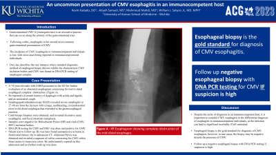
Case Description/Methods: A 74-year-old male with end stage renal disease presented to the emergency department for further evaluation of an abnormal esophagram concerning for mid to distal esophageal obstruction. This was done as he reported a 6-month history of dysphagia with solids and liquids, and an associated cough. Esophagogastroduodenoscopy (EGD) revealed severe esophagitis at 27-40 cm from the incisors with a large, nonbleeding, circumferential ulcer in the distal esophagus that extended to the gastroesophageal junction. Cold forceps biopsies were obtained, and samples underwent CMV and HSV DNA testing. Esophageal pathology revealed ulcerative acute esophagitis, and focal intestinal metaplasia. However, the samples were negative for Helicobacter Pylori and viral (CMV, HSV) inclusion bodies. DNA PCR testing for CMV and HSV were done and positive for CMV. Treatment was planned along with repeat endoscopy; however, patient was, unfortunately, lost to follow up. Three months status post EGD patient presented to the hospital in florid renal failure complicated by uremic encephalopathy and unfortunately expired. During this admission Computed Tomography abdomen and pelvis was obtained and patient was found to have a segment of colitis extending from the cecum to the transverse colon.
Discussion: Despite the rarity of diagnosis in an immunocompetent host, it is important to consider CMV esophagitis in the differential diagnosis of esophagitis in immunocompetent individuals, as the infection can lead to significant morbidity if left untreated. Esophageal biopsy is the gold standard for diagnosis of CMV esophagitis, however, in rare cases, the biopsy may be negative despite the presence of CMV. Our case demonstrates the importance of following up a negative esophageal biopsy with DNA PCR testing if suspicion is high.
Disclosures:
Kevin Kadado, DO1, Atiyeh Samadi, MS2, Mahmoud Mahdi, MD1, William J.. Salyers, MD, MPH1. P3300 - An Uncommon Presentation of CMV Esophagitis in an Immunocompetent Host, ACG 2023 Annual Scientific Meeting Abstracts. Vancouver, BC, Canada: American College of Gastroenterology.
1University of Kansas School of Medicine, Wichita, KS; 2University of Kansas-Wichita, Wichita, KS
Introduction: Gastrointestinal CMV (Cytomegalovirus) is an ulcerative process that can occur along the entirety of the gastrointestinal tract. Following colitis, esophagitis is the second most common gastrointestinal presentation of CMV. The incidence of CMV esophagitis in immunocompetent individuals is low, with most cases being reported in immunocompromised individuals. Our case outlines the rare instance of CMV esophagitis occurring in an immunocompetent individual, wherein the standard diagnostic method of esophageal biopsy did not exhibit the characteristic CMV inclusion bodies and CMV was found on DNA PCR testing of esophageal samples.
Case Description/Methods: A 74-year-old male with end stage renal disease presented to the emergency department for further evaluation of an abnormal esophagram concerning for mid to distal esophageal obstruction. This was done as he reported a 6-month history of dysphagia with solids and liquids, and an associated cough. Esophagogastroduodenoscopy (EGD) revealed severe esophagitis at 27-40 cm from the incisors with a large, nonbleeding, circumferential ulcer in the distal esophagus that extended to the gastroesophageal junction. Cold forceps biopsies were obtained, and samples underwent CMV and HSV DNA testing. Esophageal pathology revealed ulcerative acute esophagitis, and focal intestinal metaplasia. However, the samples were negative for Helicobacter Pylori and viral (CMV, HSV) inclusion bodies. DNA PCR testing for CMV and HSV were done and positive for CMV. Treatment was planned along with repeat endoscopy; however, patient was, unfortunately, lost to follow up. Three months status post EGD patient presented to the hospital in florid renal failure complicated by uremic encephalopathy and unfortunately expired. During this admission Computed Tomography abdomen and pelvis was obtained and patient was found to have a segment of colitis extending from the cecum to the transverse colon.
Discussion: Despite the rarity of diagnosis in an immunocompetent host, it is important to consider CMV esophagitis in the differential diagnosis of esophagitis in immunocompetent individuals, as the infection can lead to significant morbidity if left untreated. Esophageal biopsy is the gold standard for diagnosis of CMV esophagitis, however, in rare cases, the biopsy may be negative despite the presence of CMV. Our case demonstrates the importance of following up a negative esophageal biopsy with DNA PCR testing if suspicion is high.
Disclosures:
Kevin Kadado indicated no relevant financial relationships.
Atiyeh Samadi indicated no relevant financial relationships.
Mahmoud Mahdi indicated no relevant financial relationships.
William Salyers indicated no relevant financial relationships.
Kevin Kadado, DO1, Atiyeh Samadi, MS2, Mahmoud Mahdi, MD1, William J.. Salyers, MD, MPH1. P3300 - An Uncommon Presentation of CMV Esophagitis in an Immunocompetent Host, ACG 2023 Annual Scientific Meeting Abstracts. Vancouver, BC, Canada: American College of Gastroenterology.
