Tuesday Poster Session
Category: Esophagus
P3310 - When the Path Narrows: Navigating the Challenges of Complete Esophageal Obstruction Complicating Endoscopic Variceal Ligation
Tuesday, October 24, 2023
10:30 AM - 4:00 PM PT
Location: Exhibit Hall

Has Audio

Jordan Woodard, MD
Prisma Health
Greenville, SC
Presenting Author(s)
Jordan Woodard, MD1, Varun Moktan, MD2, Adrian Pona, MD2, Christina M. Bauer, MD, MPH2, Diego Lim, MD, MPH2
1Prisma Health, Greenville, SC; 2Prisma Health-Upstate, Greenville, SC
Introduction: Esophageal varices (EV) are a common complication of cirrhosis that have increased mortality from variceal hemorrhage if untreated. Non-selective beta blockers or endoscopic variceal ligation (EVL) are common variceal treatments. EVL can be completed at same time as variceal screening endoscopy making it convenient, especially for patients unable to tolerate beta-blocker therapy. Transient dysphagia following EVL has been described as well; although, complete esophageal obstruction is exceedingly rare with only a handful of reported cases.
Case Description/Methods: A 58-year-old male with 2-year history of Hepatocellular carcinoma and alcoholic cirrhosis complicated by esophageal varices and hypotension presented outpatient for routine variceal screening. EGD revealed grade 2 EV in the lower third of the esophagus and portal hypertensive gastropathy. EVL was performed with 2 bands placed with successful eradication and deflation of varices.
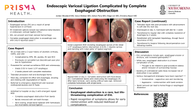
The following day the patient contacted the office with concerns regarding dysphagia, drooling and choking. He was unable to tolerate liquids or solids. Conservative management with solumedrol did not improve symptoms. Repeat EGD showed complete esophageal obstruction (Figure A). Prior bands were removed carefully with rat-tooth forceps, and single repeat band was placed with hemostasis and incomplete eradication of varices (Figure B). His symptoms rapidly resolved, and he was able to tolerate a mechanical soft diet after 2 days.
Discussion: While exceedingly rare, complete esophageal obstruction is an extremely debilitating complication of EVL requiring urgent intervention. In our case, the obstruction was likely caused by esophageal edema from banding contralateral esophageal walls. Prior reports have described management with total parenteral nutrition and bowel rest and nasogastric tube placement under direct visualization. Various techniques to remove EVL have been reported in the literature, including biopsy forceps, tip of endoscope, or loop cutters, but optimal technique is unclear. With repeat endoscopy we were able to relieve the obstruction and symptoms resolved by the following day and regular diet was manageable after 1 week. Band removal using rat-tooth forceps was both safe and effective for resolution of esophageal obstruction.

Disclosures:
Jordan Woodard, MD1, Varun Moktan, MD2, Adrian Pona, MD2, Christina M. Bauer, MD, MPH2, Diego Lim, MD, MPH2. P3310 - When the Path Narrows: Navigating the Challenges of Complete Esophageal Obstruction Complicating Endoscopic Variceal Ligation, ACG 2023 Annual Scientific Meeting Abstracts. Vancouver, BC, Canada: American College of Gastroenterology.
1Prisma Health, Greenville, SC; 2Prisma Health-Upstate, Greenville, SC
Introduction: Esophageal varices (EV) are a common complication of cirrhosis that have increased mortality from variceal hemorrhage if untreated. Non-selective beta blockers or endoscopic variceal ligation (EVL) are common variceal treatments. EVL can be completed at same time as variceal screening endoscopy making it convenient, especially for patients unable to tolerate beta-blocker therapy. Transient dysphagia following EVL has been described as well; although, complete esophageal obstruction is exceedingly rare with only a handful of reported cases.
Case Description/Methods: A 58-year-old male with 2-year history of Hepatocellular carcinoma and alcoholic cirrhosis complicated by esophageal varices and hypotension presented outpatient for routine variceal screening. EGD revealed grade 2 EV in the lower third of the esophagus and portal hypertensive gastropathy. EVL was performed with 2 bands placed with successful eradication and deflation of varices.
The following day the patient contacted the office with concerns regarding dysphagia, drooling and choking. He was unable to tolerate liquids or solids. Conservative management with solumedrol did not improve symptoms. Repeat EGD showed complete esophageal obstruction (Figure A). Prior bands were removed carefully with rat-tooth forceps, and single repeat band was placed with hemostasis and incomplete eradication of varices (Figure B). His symptoms rapidly resolved, and he was able to tolerate a mechanical soft diet after 2 days.
Discussion: While exceedingly rare, complete esophageal obstruction is an extremely debilitating complication of EVL requiring urgent intervention. In our case, the obstruction was likely caused by esophageal edema from banding contralateral esophageal walls. Prior reports have described management with total parenteral nutrition and bowel rest and nasogastric tube placement under direct visualization. Various techniques to remove EVL have been reported in the literature, including biopsy forceps, tip of endoscope, or loop cutters, but optimal technique is unclear. With repeat endoscopy we were able to relieve the obstruction and symptoms resolved by the following day and regular diet was manageable after 1 week. Band removal using rat-tooth forceps was both safe and effective for resolution of esophageal obstruction.

Figure: A. Complete esophageal obstruction noted on repeat EGD. B. Single EVL band replaced after resolution of esophageal obstruction
Disclosures:
Jordan Woodard indicated no relevant financial relationships.
Varun Moktan indicated no relevant financial relationships.
Adrian Pona indicated no relevant financial relationships.
Christina Bauer: Abbvie – Speakers Bureau. Pfizer – Advisory Committee/Board Member.
Diego Lim indicated no relevant financial relationships.
Jordan Woodard, MD1, Varun Moktan, MD2, Adrian Pona, MD2, Christina M. Bauer, MD, MPH2, Diego Lim, MD, MPH2. P3310 - When the Path Narrows: Navigating the Challenges of Complete Esophageal Obstruction Complicating Endoscopic Variceal Ligation, ACG 2023 Annual Scientific Meeting Abstracts. Vancouver, BC, Canada: American College of Gastroenterology.
