Tuesday Poster Session
Category: Esophagus
P3312 - Dysphagia Caused by a Rare Case of Esophageal
Tuesday, October 24, 2023
10:30 AM - 4:00 PM PT
Location: Exhibit Hall

Has Audio

Marisa Pope, DO
Jefferson Health
Stratford, NJ
Presenting Author(s)
Marisa Pope, DO1, Joshua Soliman, DO2, Lucy Joo, DO3
1Jefferson Health, Stratford, NJ; 2Thomas Jefferson University, Voorhees, NJ; 3Jefferson Health, Cherry Hill, NJ
Introduction: Neuroendocrine tumors are derived from endocrine cells that comprise only ~1% of cells in the gut and pancreas. These cells comprise both the intestinal crypts and islets of Langerhans. Of all neuroendocrine tumors diagnosed annually only ~1.6% of them are found in the esophagus. The most common locations for gastroenteropancreatic neuroendocrine tumors are within the ileum, rectum, and appendix. There are few case reports describing esophageal neuroendocrine tumors. We present a case of a neuroendocrine esophageal tumor presenting as dysphagia resulting in early diagnosis.
Case Description/Methods: A 59-year-old female with a significant past medical history of tobacco use, alcohol use, HTN, and depression presented to the emergency room with emesis. Over a period of several months, she began experiencing emesis after every meal. She was unable to eat foods of certain textures and endorsed a sternal sensation. She was able to tolerate liquids without issue. She admitted to drinking multiple beers daily and tobacco use. She had unintentionally lost over twenty pounds over a 3 month period. She had a CT chest abdomen, and pelvis that showed a distal esophageal mass with fluid distension in the more proximal esophagus with associated gastrohepatic nodal metastasis. The patient was seen by gastroenterology and underwent an esophageal endoscopy and ultrasound. A biopsy of the esophageal mass was consistent with a neuroendocrine tumor of the esophagus. It showed fragments of squamous mucosa with underlying poorly differentiated carcinoma with neuroendocrine features. She was started on systemic chemotherapy, including Leucovorin, Oxaliplatin, and Fluorouracil.
Discussion: Esophageal neuroendocrine tumors only represent about 0.03-0.05% of all known esophageal cancers. The presentation of this tumor is variable as some patients are asymptomatic and others experience dysphagia and weight loss as early symptoms, such as our patient. These early symptoms facilitated prompt intervention by undergoing an endoscopic ultrasound and biopsy evaluation leading to earlier diagnosis and better prognosis in this patient. An exact standard of treatment has not been established, but typically these patients undergo combination therapy of radiation, chemotherapy, and surgical excision. Due to the rarity of reports of esophageal neuroendocrine tumors ours help add to our understanding of early recognition. Further, enforcing the importance of esophageal endoscopy in patients that develop new-onset dysphagia.

Disclosures:
Marisa Pope, DO1, Joshua Soliman, DO2, Lucy Joo, DO3. P3312 - Dysphagia Caused by a Rare Case of Esophageal, ACG 2023 Annual Scientific Meeting Abstracts. Vancouver, BC, Canada: American College of Gastroenterology.
1Jefferson Health, Stratford, NJ; 2Thomas Jefferson University, Voorhees, NJ; 3Jefferson Health, Cherry Hill, NJ
Introduction: Neuroendocrine tumors are derived from endocrine cells that comprise only ~1% of cells in the gut and pancreas. These cells comprise both the intestinal crypts and islets of Langerhans. Of all neuroendocrine tumors diagnosed annually only ~1.6% of them are found in the esophagus. The most common locations for gastroenteropancreatic neuroendocrine tumors are within the ileum, rectum, and appendix. There are few case reports describing esophageal neuroendocrine tumors. We present a case of a neuroendocrine esophageal tumor presenting as dysphagia resulting in early diagnosis.
Case Description/Methods: A 59-year-old female with a significant past medical history of tobacco use, alcohol use, HTN, and depression presented to the emergency room with emesis. Over a period of several months, she began experiencing emesis after every meal. She was unable to eat foods of certain textures and endorsed a sternal sensation. She was able to tolerate liquids without issue. She admitted to drinking multiple beers daily and tobacco use. She had unintentionally lost over twenty pounds over a 3 month period. She had a CT chest abdomen, and pelvis that showed a distal esophageal mass with fluid distension in the more proximal esophagus with associated gastrohepatic nodal metastasis. The patient was seen by gastroenterology and underwent an esophageal endoscopy and ultrasound. A biopsy of the esophageal mass was consistent with a neuroendocrine tumor of the esophagus. It showed fragments of squamous mucosa with underlying poorly differentiated carcinoma with neuroendocrine features. She was started on systemic chemotherapy, including Leucovorin, Oxaliplatin, and Fluorouracil.
Discussion: Esophageal neuroendocrine tumors only represent about 0.03-0.05% of all known esophageal cancers. The presentation of this tumor is variable as some patients are asymptomatic and others experience dysphagia and weight loss as early symptoms, such as our patient. These early symptoms facilitated prompt intervention by undergoing an endoscopic ultrasound and biopsy evaluation leading to earlier diagnosis and better prognosis in this patient. An exact standard of treatment has not been established, but typically these patients undergo combination therapy of radiation, chemotherapy, and surgical excision. Due to the rarity of reports of esophageal neuroendocrine tumors ours help add to our understanding of early recognition. Further, enforcing the importance of esophageal endoscopy in patients that develop new-onset dysphagia.

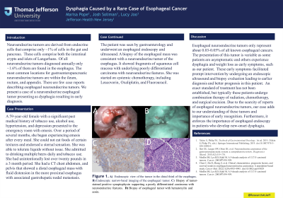
Figure: The top images are two endoscopic views of the esophageal neuroendocrine tumor.
The two bottom images are hematoxylin-eosn stain on the right (400x) showing tumor cells with scant cytoplasm, small round to oval nuclei with dispersed chromatin, inconspicuous nucleoli and frequent mitoses. The image on the bottom left shows an immunohistochemical stain for synaptophysin (400x).
The two bottom images are hematoxylin-eosn stain on the right (400x) showing tumor cells with scant cytoplasm, small round to oval nuclei with dispersed chromatin, inconspicuous nucleoli and frequent mitoses. The image on the bottom left shows an immunohistochemical stain for synaptophysin (400x).
Disclosures:
Marisa Pope indicated no relevant financial relationships.
Joshua Soliman indicated no relevant financial relationships.
Lucy Joo indicated no relevant financial relationships.
Marisa Pope, DO1, Joshua Soliman, DO2, Lucy Joo, DO3. P3312 - Dysphagia Caused by a Rare Case of Esophageal, ACG 2023 Annual Scientific Meeting Abstracts. Vancouver, BC, Canada: American College of Gastroenterology.
