Tuesday Poster Session
Category: Esophagus
P3318 - A Rare Case of Esophageal Epidermoid Metaplasia and Esophageal Candidiasis
Tuesday, October 24, 2023
10:30 AM - 4:00 PM PT
Location: Exhibit Hall

Has Audio

Aya Tabbalat, MD
Case Western Reserve University - University Hospitals Cleveland Medical Center
Cleveland, OH
Presenting Author(s)
Mohammed Z. Sheriff, MD1, Amitabh Chak, MD2, Alwalid Ammoun, MD3, Aya Tabbalat, MD1
1Case Western Reserve University - University Hospitals Cleveland Medical Center, Cleveland, OH; 2Digestive Health Institute, University Hospitals Cleveland Medical Center, Cleveland, OH; 3University Hospitals Cleveland Medical Center-Case Western Reserve University, Cleveland, OH
Introduction: Esophageal epidermoid metaplasia (EEM) is a rare premalignant condition seen in middle aged men with history of alcohol and tobacco use. EEM has been reported in association with esophageal squamous cell carcinoma, as well as GERD, Barrett’s esophagus, lichen planus, and esophageal adenocarcinoma.
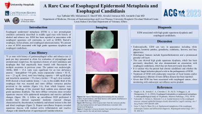
Case Description/Methods: A 75 yo man with history of gastroesophageal reflux and tobacco use (1 pack per day) presented to clinic for evaluation of odynophagia and unintentional weight loss. He reported a history of oral Candidiasis and dysphagia that had empirically been treated with fluconazole on multiple occasions in previous years. The patient was underweight, 104 lbs, BMI = 15.4. Labs were significant for an iron deficiency anemia – hemoglobin= 9.0 g/dL, mean corpuscular volume = 82 fL, iron = 23 ug/dL (low), total iron binding capacity = 469 ug/dL(high) and transferrin saturation = 5% (low), ferritin = 59 ug/L (low normal). EGD showed a focal nodular lesion,< 1 cm, in the middle third of the esophagus that was resected and more diffuse verrucous patches proximal to the nodules (figure 1A), from which biopsies were obtained. Histology of the resected focal nodular area showed high grade squamous dysplasia. The more diffuse verrucous areas revealed squamous mucosa with epidermoid metaplasia and reactive epithelial changes (figure 1B). A follow up surveillance EGD performed 4 months later showed segmental moderate mucosal changes characterized by discoloration, nodularity and altered texture in the mid and distal esophagus (figure 2). Repeat surveillance biopsies revealed squamous mucosa with marked active inflammation and reactive changes with identification of superimposed Candida.
Discussion: Endoscopically, EEM can vary in appearance including white plaques, keratotic patches,granularity, nodularity, furrows, and lacy appearance. Histological features include hyperorthokeratosis and a pronounced granular cell layer. This case showed high grade squamous dysplasia, which has been previously described but also demonstrated as association with esophageal Candidiasis, which has not been previously described. It is unclear why the patient had chronic Candidiasis and whether the chronic fungal infection played a role in the pathogenesis of EEM. Treatment of EEM with endoscopic resection of focal lesions and/or radiofrequency ablation of more diffuse disease has been reported. Given that EEM has been associated with esophageal cancer, close monitoring with endoscopic surveillance seems prudent.

Disclosures:
Mohammed Z. Sheriff, MD1, Amitabh Chak, MD2, Alwalid Ammoun, MD3, Aya Tabbalat, MD1. P3318 - A Rare Case of Esophageal Epidermoid Metaplasia and Esophageal Candidiasis, ACG 2023 Annual Scientific Meeting Abstracts. Vancouver, BC, Canada: American College of Gastroenterology.
1Case Western Reserve University - University Hospitals Cleveland Medical Center, Cleveland, OH; 2Digestive Health Institute, University Hospitals Cleveland Medical Center, Cleveland, OH; 3University Hospitals Cleveland Medical Center-Case Western Reserve University, Cleveland, OH
Introduction: Esophageal epidermoid metaplasia (EEM) is a rare premalignant condition seen in middle aged men with history of alcohol and tobacco use. EEM has been reported in association with esophageal squamous cell carcinoma, as well as GERD, Barrett’s esophagus, lichen planus, and esophageal adenocarcinoma.
Case Description/Methods: A 75 yo man with history of gastroesophageal reflux and tobacco use (1 pack per day) presented to clinic for evaluation of odynophagia and unintentional weight loss. He reported a history of oral Candidiasis and dysphagia that had empirically been treated with fluconazole on multiple occasions in previous years. The patient was underweight, 104 lbs, BMI = 15.4. Labs were significant for an iron deficiency anemia – hemoglobin= 9.0 g/dL, mean corpuscular volume = 82 fL, iron = 23 ug/dL (low), total iron binding capacity = 469 ug/dL(high) and transferrin saturation = 5% (low), ferritin = 59 ug/L (low normal). EGD showed a focal nodular lesion,< 1 cm, in the middle third of the esophagus that was resected and more diffuse verrucous patches proximal to the nodules (figure 1A), from which biopsies were obtained. Histology of the resected focal nodular area showed high grade squamous dysplasia. The more diffuse verrucous areas revealed squamous mucosa with epidermoid metaplasia and reactive epithelial changes (figure 1B). A follow up surveillance EGD performed 4 months later showed segmental moderate mucosal changes characterized by discoloration, nodularity and altered texture in the mid and distal esophagus (figure 2). Repeat surveillance biopsies revealed squamous mucosa with marked active inflammation and reactive changes with identification of superimposed Candida.
Discussion: Endoscopically, EEM can vary in appearance including white plaques, keratotic patches,granularity, nodularity, furrows, and lacy appearance. Histological features include hyperorthokeratosis and a pronounced granular cell layer. This case showed high grade squamous dysplasia, which has been previously described but also demonstrated as association with esophageal Candidiasis, which has not been previously described. It is unclear why the patient had chronic Candidiasis and whether the chronic fungal infection played a role in the pathogenesis of EEM. Treatment of EEM with endoscopic resection of focal lesions and/or radiofrequency ablation of more diffuse disease has been reported. Given that EEM has been associated with esophageal cancer, close monitoring with endoscopic surveillance seems prudent.

Figure: Figure 1A: Narrow Band Imaging demonstration few tiny mucosal nodules, hyper-keratosis, and linear furrows with a patchy distribution in the middle third of the esophagus.
Figure 1B: Histology of the biopsies taken from the verrucous patches showing hypergranulosis and hyperorthokeratosis consistent with Epidermoid Metaplasia.
Figure 2: Segmental verrucous keratotic focal and broad linear mucosal changes noted in the mid and distal esophagus.
Figure 1B: Histology of the biopsies taken from the verrucous patches showing hypergranulosis and hyperorthokeratosis consistent with Epidermoid Metaplasia.
Figure 2: Segmental verrucous keratotic focal and broad linear mucosal changes noted in the mid and distal esophagus.
Disclosures:
Mohammed Sheriff indicated no relevant financial relationships.
Amitabh Chak indicated no relevant financial relationships.
Alwalid Ammoun indicated no relevant financial relationships.
Aya Tabbalat indicated no relevant financial relationships.
Mohammed Z. Sheriff, MD1, Amitabh Chak, MD2, Alwalid Ammoun, MD3, Aya Tabbalat, MD1. P3318 - A Rare Case of Esophageal Epidermoid Metaplasia and Esophageal Candidiasis, ACG 2023 Annual Scientific Meeting Abstracts. Vancouver, BC, Canada: American College of Gastroenterology.
